Comparative Study
. 2007 Jan;75(1):35-43.
doi: 10.1128/IAI.00963-06. Epub 2006 Oct 23.

Analysis of Bartonella adhesin A expression reveals differences between various B. henselae strains

Affiliations
Comparative Study

Analysis of Bartonella adhesin A expression reveals differences between various B. henselae strains

Tanja Riess et al. Infect Immun. 2007 Jan.

Abstract

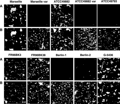
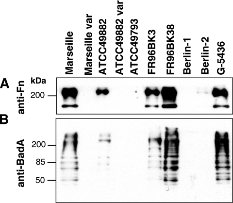
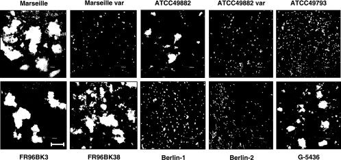
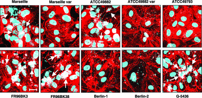
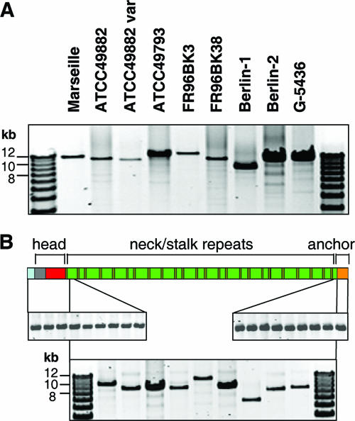
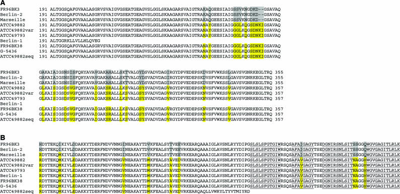
Bartonella henselae causes cat scratch disease and the vasculoproliferative disorders bacillary angiomatosis and peliosis hepatis in humans. One of the best known pathogenicity factors of B. henselae is Bartonella adhesin A (BadA), which is modularly constructed, consisting of head, neck/stalk, and membrane anchor domains. BadA is important for the adhesion of B. henselae to extracellular-matrix proteins and endothelial cells (ECs). In this study, we analyzed different B. henselae strains for BadA expression, autoagglutination, fibronectin (Fn) binding, and adhesion to ECs. We found that the B. henselae strains Marseille, ATCC 49882, Freiburg 96BK3 (FR96BK3), FR96BK38, and G-5436 express BadA. Remarkably, BadA expression was lacking in a B. henselae ATCC 49882 variant, in strains ATCC 49793 and Berlin-1, and in the majority of bacteria of strain Berlin-2. Adherence of B. henselae to ECs and Fn reliably correlated with BadA expression. badA was present in all tested strains, although the length of the gene varied significantly due to length variations of the stalk region. Sequencing of the promoter, head, and membrane anchor regions revealed only minor differences that did not correlate with BadA expression, apart from strain Berlin-1, in which a 1-bp deletion led to a frameshift in the head region of BadA. Our data suggest that, apart from the identified genetic modifications (frameshift deletion and recombination), other so-far-unknown regulatory mechanisms influence BadA expression. Because of variations between and within different B. henselae isolates, BadA expression should be analyzed before performing infection experiments with B. henselae.


Figures

FIG. 1.
FIG. 1.
BadA expression in different B. henselae strains. (A) BadA was detected on the surfaces of the strains Marseille, ATCC 49882, FR96BK3, FR96BK38, and G-5436 by immunofluorescence using a specific anti-BadA-Ab (var., variant strain). Note that in strain Berlin-2, only a few bacteria are BadA positive. (B) For internal control, bacterial DNA was stained with DAPI. Scale bar, 8 μm.
FIG. 2.
FIG. 2.
Analysis of the Fn-binding capacity of B. henselae and detection of BadA expression by Western blotting. (A) Fn (240 kDa) bound to bacteria was detected in bacterial lysates using an anti-Fn Ab. Note that only BadA-positive strains bind Fn. Strain Berlin-2, in which only a few bacteria express BadA (see Fig. 1), shows very weak Fn binding. (B) For internal control, BadA (∼340 kDa) was detected in bacterial lysates by using a specific anti-BadA Ab. Multiple bands are interpreted as degradation products of the high-molecular-weight BadA (29).
FIG. 3.
FIG. 3.
BadA-dependent autoagglutination of different B. henselae strains. Bacteria were resuspended in PBS, incubated for 60 min, stained with DAPI, and analyzed by CLSM. Note that only BadA-positive strains show autoagglutination. Scale bar, 20 μm.
FIG. 4.
FIG. 4.
BadA-dependent adhesion of different B. henselae strains to endothelial cells. HUVECs were infected with B. henselae, and adhesion of the bacteria was analyzed after 30 min by CLSM. Bacteria and host cell nuclei were stained with DAPI (blue signal); filamentous actin was stained with TRITC-labeled phalloidin (red signal). Note that only BadA-positive strains adhere strongly to HUVECs (arrows). Scale bar, 20 μm.
FIG. 5.
FIG. 5.
Presence and lengths of badA, badAhead, badAstalk, and badAmembrane anchor of different B. henselae strains analyzed by PCR. (A) Long-distance PCRs were performed (for details, see Materials and Methods), and the PCR products were analyzed in a 0.6% agarose gel. All B. henselae strains harbor badA, although there are differences in the size of the gene (∼9 to 12 kb). (B) Detection of the badAhead- (upper left), badAstalk- (lower middle) and badAmembrane anchor-coding sequences (upper right) in different B. henselae isolates. Note that differences in size are detectable only in badAstalk. For reasons of clarity, BadA is schematically depicted with the signal peptide (light blue), the head sequence (gray and red), 24 neck/stalk repeats (brown and green, respectively) and the membrane anchor (orange).
FIG. 6.
FIG. 6.
Comparison of the head and membrane anchor regions of BadA in different B. henselae strains. A Clustal W alignment of (A) amino acids 191 to 357 (part of the head) and (B) the C-terminal 130 amino acids (membrane anchor) is shown. Amino acids 1 to 190 are not shown, as they are identical in all strains. Differences in the sequences are marked in yellow and gray. ATCC49882seq, sequence of the published ATCC 49882 isolate (1). In this strain, a stop codon is introduced in the membrane anchor-coding sequence due to a 1-bp deletion. The strains cluster, in both the head and the membrane anchor regions, in two groups, with B. henselae Marseille as an intermediate between the groups. The four transmembrane β-strands of the membrane anchor are underlined.

Similar articles

Cited by

References

    1. Alsmark, C. M., A. C. Frank, E. O. Karlberg, B. A. Legault, D. H. Ardell, B. Canback, A. S. Eriksson, A. K. Naslund, S. A. Handley, M. Huvet, B. LaScola, M. Holmberg, and S. G. Andersson. 2004. The louse-borne human pathogen Bartonella quintana is a genomic derivative of the zoonotic agent Bartonella henselae. Proc. Natl. Acad. Sci. USA 101:9716-9721. - PMC - PubMed
    1. Anderson, B. E., and M. A. Neuman. 1997. Bartonella spp. as emerging human pathogens. Clin. Microbiol. Rev. 10:203-219. - PMC - PubMed
    1. Arvand, M., C. Wendt, T. Regnath, R. Ullrich, and H. Hahn. 1998. Characterization of Bartonella henselae isolated from bacillary angiomatosis lesions in a human immunodeficiency virus-infected patient in Germany. Clin. Infect. Dis. 26:1296-1299. - PubMed
    1. Batterman, H. J., J. A. Peek, J. S. Loutit, S. Falkow, and L. S. Tompkins. 1995. Bartonella henselae and Bartonella quintana adherence to and entry into cultured human epithelial cells. Infect. Immun. 63:4553-4556. - PMC - PubMed
    1. Centers for Disease Control and Prevention. 1999. Serodiagnosis of emerging infectious diseases: Bartonella and Ehrlichia infections. Centers for Disease Control and Prevention, Manassas, VA.

Publication types

MeSH terms

LinkOut - more resources