. 2007 Jan;75(1):201-10.
doi: 10.1128/IAI.01327-06. Epub 2006 Oct 23.

Role of monocyte-acquired hemozoin in suppression of macrophage migration inhibitory factor in children with severe malarial anemia

Affiliations

Role of monocyte-acquired hemozoin in suppression of macrophage migration inhibitory factor in children with severe malarial anemia

Gordon A Awandare et al. Infect Immun. 2007 Jan.

Abstract

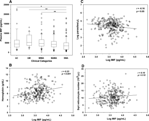
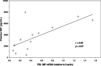
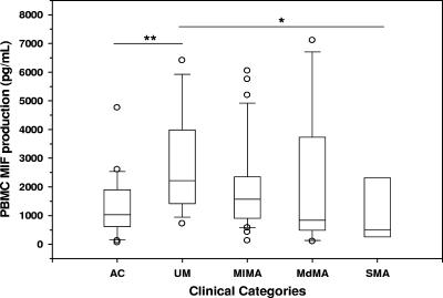
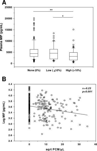
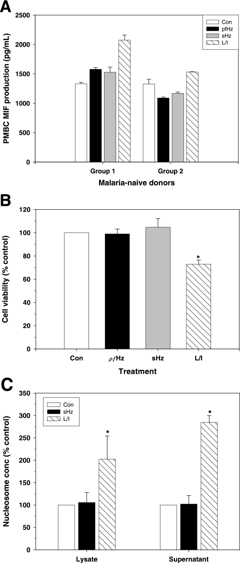
Severe malarial anemia (SMA), caused by Plasmodium falciparum infections, is one of the leading causes of childhood mortality in sub-Saharan Africa. Although the molecular determinants of SMA are largely undefined, dysregulation in host-derived inflammatory mediators influences disease severity. Macrophage migration inhibitory factor (MIF) is an important regulator of innate inflammatory responses that has recently been shown to suppress erythropoiesis and promote pathogenesis of SMA in murine models. To examine the role of MIF in the development of childhood SMA, peripheral blood MIF production was examined in Kenyan children (aged <3 years, n = 357) with P. falciparum malarial anemia. All children in the study were free from bacteremia and human immunodeficiency virus type 1. Since deposition of malarial pigment (hemozoin [Hz]) contributes to suppression of erythropoiesis, the relationship between MIF concentrations and monocytic acquisition of Hz was also examined in vivo and in vitro. Circulating MIF concentrations declined with increasing severity of anemia and significantly correlated with peripheral blood leukocyte MIF transcripts. However, MIF concentrations in peripheral blood were not significantly associated with reticulocyte production. Multivariate regression analyses, controlling for age, gender, and parasitemia, further revealed that elevated levels of pigment-containing monocytes (PCM) was associated with SMA and decreased MIF production. In addition, PCM levels were a better predictor of hemoglobin and MIF concentrations than parasite density. Additional experiments in malaria-naive individuals demonstrated that hemozoin caused both increased and decreased MIF production in cultured peripheral blood mononuclear cells (PBMC) in a donor-specific manner, independent of apoptosis. However, PBMC MIF production in children with acute malaria progressively declined with increasing anemia severity. Results presented here demonstrate that acquisition of hemozoin by monocytes is associated with suppression of peripheral blood MIF production and enhanced severity of anemia in childhood malaria.


Figures

FIG. 1.
FIG. 1.
Relationship of plasma MIF with anemia, parasite density, and absolute reticulocyte number. Plasma levels of MIF in children with malaria and controls were determined by ELISA. (A) Data are presented according to the following anemia categories: UM (Hb ≥ 11.0 g/dl, n = 23), MlMA (8.0 ≤ Hb < 11.0 g/dl, n = 71), MdMA (6.0 ≤ Hb < 8.0 g/dl, n = 94), and SMA (Hb < 6.0 g/dl, n = 109). AC (n = 39) with Hb levels of ≥11.0 g/dl were used as a reference group. Boxes represent the interquartile range, the line through the box is the median, whiskers show 10th and 90th percentiles, and symbols are outliers. Median (interquartile range) levels of MIF were as follows (in pg/ml): AC, 4,383 (2,807 to 6,376); UM, 5,225 (3,615 to 9,071); MlMA, 4,611 (3,270 to 6,665); MdMA, 4,197 (2,862 to 5,743); SMA, 3,422 (1,566 to 4,993). *, P < 0.05; **, P < 0.001. Mann-Whitney U tests conducted after analysis of variance (Kruskal-Wallis test) revealed significant differences across groups. Linear relationships of plasma MIF levels with hemoglobin concentrations (B), parasite density (C), and ARN (D) in children with malaria (n = 298) are shown as scatter plots. MIF concentrations and parasitemia were log transformed, while the ARN was square root (sqrt) transformed for normality. Statistical associations were determined by Pearson's correlation tests.
FIG. 2.
FIG. 2.
Correlation of PBL MIF mRNA with circulating MIF. Levels of MIF mRNA in PBL (n = 13) were determined by real-time reverse transcription-PCR and expressed relative to endogenous β-actin mRNA levels. The scatter plot shows the correlation of PBL MIF mRNA with circulating MIF levels in matched samples. Statistical association was determined by Pearson's rank correlation test.
FIG. 3.
FIG. 3.
MIF production from PBMC of children with acute malaria. PBMC were isolated from the following groups of children with acute malaria and healthy controls (AC) and cultured for 48 h: AC (n = 24), UM (n = 15), MlMA (n = 37), MdMA (n = 13), and SMA (n = 5). MIF concentrations in culture supernatants were determined by ELISA and presented as box plots in which the box represents the interquartile range, the line through the box is the median, whiskers show 10th and 90th percentiles, and symbols are outliers. Median (interquartile range) levels of MIF were as follows (in pg/ml): AC, 1,034 (615 to 1,902); UM, 2,218 (1,418 to 3,989); MlMA, 1,609 (940 to 2,439); MdMA, 842 (495 to 3,737); SMA, 511 (264 to 2,312). *, P < 0.05; **, P < 0.005. Mann-Whitney U tests conducted after analysis of variance (Kruskal-Wallis test) revealed significant differences between the groups.
FIG. 4.
FIG. 4.
Relationship between plasma MIF levels and pigment-containing monocytes. Giemsa-stained blood smears obtained from children with malaria were examined for the presence of PCM. (A) Plasma levels of MIF are presented according to percentage of total monocytes containing pigment, as follows: none (no PCM observed, n = 149), low (≤10% PCM, n = 60), and high (>10% PCM, n = 79). Boxes represent the interquartile range, the line through the box is the median, whiskers show 10th and 90th percentiles, and symbols are outliers. Median (interquartile range) levels of MIF were as follows (in pg/ml): no PCM, 4,417 (3,112 to 6,266); low PCM, 4,584 (2,019 to 6,331); high PCM, 4,417 (3,112 to 6,266). *, P < 0.05; **, P < 0.005. Mann-Whitney U tests conducted after analysis of variance (Kruskal-Wallis test) revealed significant differences across groups. (B) Correlation between circulating MIF concentrations and PCM/μl of blood. MIF levels were log transformed, while PCM/μl values were square root (sqrt) transformed for normality, and Pearson's correlation test was used to examine statistical association.
FIG. 5.
FIG. 5.
Effects of hemozoin on MIF production and apoptosis of cultured leukocytes. PBMC from malaria-naive donors were cultured (1 × 10⁶ cells/ml) in the presence of media alone (Con), 10 μg/ml hemozoin (pfHz), 10 μg/ml β-hematin (sHz), or a combination of 100 ng/ml LPS (L) and 200 U/ml IFN-γ (I). (A) Supernatants were harvested after 48 h of incubation, and MIF concentrations were determined by ELISA. Data shown are for three donors representative of individuals for whom MIF increased (group 1) and three donors representative of individuals for whom MIF decreased (group 2) in response to stimulation with pfHz and sHz. Differences in MIF levels between treated cells and control conditions were statistically significant (P < 0.05 for all comparisons, Student's t test). (B) Cell viability was assessed in PBMC from 3 donors after 48 h of culture using a MTT assay and expressed as a percentage of the control result. (C) PBMC apoptosis was determined by detecting nucleosomes in cell lysates and supernatants at 48 h using ELISA. Data are presented as means (±standard errors of the means) of results from independent experiments. *, P < 0.05 compared to Con, Student's t test.

References

    1. Abdalla, S. H. 1990. Hematopoiesis in human malaria. Blood Cells 16:401-419. - PubMed
    1. Akpede, G. O., and R. M. Sykes. 1993. Malaria with bacteraemia in acutely febrile preschool children without localizing signs: coincidence or association/complication? J. Trop. Med. Hyg. 96:146-150. - PubMed
    1. Alourfi, Z., R. P. Donn, A. Stevens, A. Berry, A. McMaster, and D. W. Ray. 2005. Glucocorticoids suppress macrophage migration inhibitory factor (MIF) expression in a cell-type-specific manner. J. Mol. Endocrinol. 34:583-595. - PubMed
    1. Amodu, O. K., A. A. Adeyemo, P. E. Olumese, and R. A. Gbadegesin. 1998. Intraleucocytic malaria pigment and clinical severity of malaria in children. Trans. R. Soc. Trop. Med. Hyg. 92:54-56. - PubMed
    1. Arese, P., and E. Schwarzer. 1997. Malarial pigment (haemozoin): a very active ‘inert’ substance. Ann. Trop. Med. Parasitol. 91:501-516. - PubMed

Publication types