. 2006 Oct 30;203(11):2433-40.
doi: 10.1084/jem.20061302. Epub 2006 Oct 23.

Signal-dependent splicing of tissue factor pre-mRNA modulates the thrombogenicity of human platelets

Affiliations

Signal-dependent splicing of tissue factor pre-mRNA modulates the thrombogenicity of human platelets

Hansjörg Schwertz et al. J Exp Med. .

Abstract

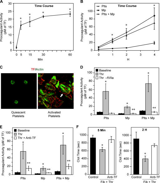
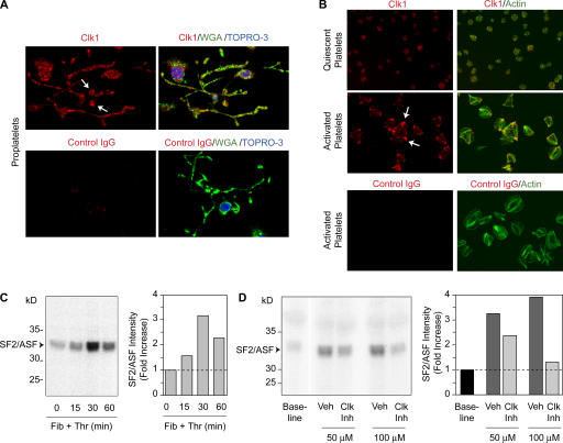
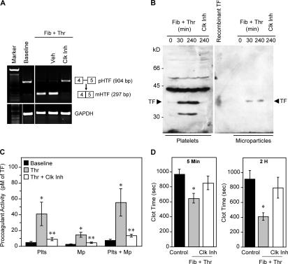
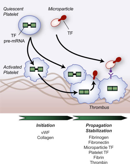
Tissue factor (TF) is an essential cofactor for the activation of blood coagulation in vivo. We now report that quiescent human platelets express TF pre-mRNA and, in response to activation, splice this intronic-rich message into mature mRNA. Splicing of TF pre-mRNA is associated with increased TF protein expression, procoagulant activity, and accelerated formation of clots. Pre-mRNA splicing is controlled by Cdc2-like kinase (Clk)1, and interruption of Clk1 signaling prevents TF from accumulating in activated platelets. Elevated intravascular TF has been reported in a variety of prothrombotic diseases, but there is debate as to whether anucleate platelets-the key cellular effector of thrombosis-express TF. Our studies demonstrate that human platelets use Clk1-dependent splicing pathways to generate TF protein in response to cellular activation. We propose that platelet-derived TF contributes to the propagation and stabilization of a thrombus.


Figures

Figure 1.
Figure 1.
Platelets contain TF pre-mRNA and splice it into mature message in response to activation. (A) TF and GAPDH mRNA expression in freshly isolated platelets (0 min), platelets adherent to fibrinogen and coactivated with thrombin (Fib + Thr), or HL60 leukocytes. The negative control lane (Neg con) is a PCR reaction conducted without cDNA template. A is representative of two independent experiments. (B and C) TF and GAPDH mRNA expression in freshly isolated platelets (Control) or platelets that are adherent to fibrinogen in the presence of thrombin (Fib + Thr). The negative control lane (Neg con) in C is a PCR reaction conducted without cDNA template. B and C are representative of >10 independent experiments. (D) Indirect in situ PCR for intronic TF pre-mRNA in megakaryocytes (left) and megakaryocytes with proplatelet extensions (right). In the bottom panels (no RT), the reverse transcriptase (RT) was omitted from the RT reaction. (E) Direct in situ PCR for TF pre-mRNA was conducted in quiescent platelets (top left), whereas PCR for mature TF mRNA was conducted in platelets adherent to fibrinogen in the presence of thrombin for 1 h (top right). In the bottom panels (no RT), the reverse transcriptase was omitted during the RT reaction. D and E are representative of three independent experiments.
Figure 2.
Figure 2.
Platelet activation increases TF-dependent procoagulant activity. (A and B) A timecourse (A, 0–60 min; B, 0–4 h) of TF-dependent procoagulant activity in platelets that have adhered to fibrinogen in the presence of thrombin. In B, each line represents procoagulant activity from platelet membranes (Plts), platelet-derived microparticles (Mp), or platelet membranes together with microparticles from the same sample (Plts + Mp). The data are displayed as pM of TF per 2 × 10⁹ platelets and represent the mean ± SEM of three independent experiments. (C) Immunolocalization of TF in freshly isolated platelets (left) and platelets that have adhered and spread on immobilized fibrinogen in the presence of thrombin for 2 h (right). The green stain in both panels represents actin. The red stain in the right panel shows immunolocalization of TF on the surface of activated platelets (see arrow). This experiment is representative of two independent studies. (D and E) The bars (n = 4) in these panels show TF-dependent procoagulant activity in freshly isolated platelets or platelets activated as in B for 4 h in the presence or absence of factor VIIa (D) or a neutralizing antibody directed against TF (E). A single asterisk (*) indicates a statistically significant difference (P < 0.05) in TF-dependent procoagulant activity between freshly isolated (baseline) and activated platelets; the double asterisk (**) represents a significant difference (P < 0.05) between activated platelets under untreated or treated conditions. (F) These panels show clot formation in plasma that is incubated with membranes isolated from quiescent platelets or platelets activated with fibrinogen and thrombin for 5 min (left) or 2 h (right). The white bars represent activated platelets that were incubated with a neutralizing antibody directed against tissue factor (Anti-TF). The data represent the mean ± SEM of six independent experiments.
Figure 3.
Figure 3.
Platelet Clk1 induces SF2/ASF phosphorylation. (A) Megakaryocytes with proplatelet extensions were stained with anti-Clk1 (top) or control IgG (bottom). The right panels are overlays where anti-Clk1 or IgG are in red, wheat germ agglutinin is in green, and topro-3, which stains nuclei, is in blue. (B) Platelets were left quiescent (top) or activated by adherence to immobilized fibrinogen in the presence of thrombin for 1 h (middle and bottom). The cells were stained with anti-Clk1 (top and middle) or control IgG (bottom). The right panels are overlays in which anti-Clk1 or IgG is in red, polymerized actin is in green, and colocalization of the two markers is identified by yellow staining. A and B are representative images of multiple cells taken from three and four independent experiments, respectively. (C and D) Platelets were activated as in B, lysed, and immune complex kinase assays for Clk1 activity were performed with recombinant SF2/ASF as the substrate. C shows a time course of Clk1-dependent SF2/ASF phosphorylation in activated platelet lysates. D shows Clk1-dependent SF2/ASF phosphorylation in freshly isolated platelet lysates or platelets that were pretreated with vehicle or Tg003 (Clk Inh) (lanes 2–5). The bars for C and D represent fold increases in SF2/ASF phosphorylation over baseline as estimated by densitometry. The gels in C and D are representative of three independent experiments.
Figure 4.
Figure 4.
Interruption of Clk1 blocks signal-dependent TF pre-mRNA splicing and bioactive protein accumulation in platelets. (A) Human TF pre-mRNA (pHTF; 904 bp) and mature mRNA (mHTF; 297 bp) was determined in freshly isolated platelets and in platelets adherent to immobilized fibrinogen and coactivated with thrombin for 2 h. The activated platelets were either left untreated (lane 3) or pretreated with vehicle (Veh) or Tg003 (Clk Inh). (B) Western blot analysis showing TF protein expression in platelets and platelet-derived microparticles. The platelets were left quiescent (baseline) or adhered to immobilized fibrinogen and coactivated with thrombin for 30 min or 4 h in the presence or absence of Tg003 (Clk Inh). Recombinant TF was used as a positive control. The panels in A and B are representative of at least three independent experiments. (C) This panel depicts TF-dependent procoagulant activity in freshly isolated platelets or platelets activated, as in Fig. 2 B, in the presence or absence of the Clk inhibitor. The data are displayed as pM of TF per 2 × 10⁹ platelets and represent the mean ± SEM of six independent experiments. A single asterisk (*) indicates a statistically significant difference (P < 0.05) in TF-dependent procoagulant activity between freshly isolated (baseline) and activated platelets; the double asterisk (**) represents a significant difference (P < 0.05) between activated platelets under untreated or treated conditions. (D) These panels show plasma clot formation in the presence of membranes isolated from freshly isolated platelets (Control) or platelets activated for 5 min (left) or 2 h (right) with fibrinogen and thrombin. The activated platelets were pretreated with either the Clk inhibitor (Clk Inh) or DMSO. The data represent the mean ± SEM of five independent experiments. The single asterisk (*) indicates a statistically significant difference (P < 0.05) in the rate of clot formation in plasma samples exposed to fibrinogen and thrombin-treated platelets compared with quiescent platelets (Control) and activated platelets treated with the Clk inhibitor.
Figure 5.
Figure 5.
Proposed model by which platelet-derived tissue factor contributes to propagation and stabilization of a thrombus.

References

    1. Jurk, K., and B.E. Kehrel. 2005. Platelets: physiology and biochemistry. Semin. Thromb. Hemost. 31:381–392. - PubMed
    1. Ruggeri, Z.M. 2002. Platelets in atherothrombosis. Nat. Med. 8:1227–1234. - PubMed
    1. Roberts, H.R., M. Hoffman, and D.M. Monroe. 2006. A cell-based model of thrombin generation. Semin. Thromb. Hemost. 32(Suppl 1):32–38. - PubMed
    1. Lentz, B.R. 2003. Exposure of platelet membrane phosphatidylserine regulates blood coagulation. Prog. Lipid Res. 42:423–438. - PubMed
    1. Morrissey, J.H., P.F. Neuenschwander, Q. Huang, C.D. McCallum, B. Su, and A.E. Johnson. 1997. Factor VIIa-tissue factor: functional importance of protein-membrane interactions. Thromb. Haemost. 78:112–116. - PubMed

Publication types

MeSH terms