. 2006 Oct 23;175(2):209-15.
doi: 10.1083/jcb.200606016.

Toll-like receptor 8 functions as a negative regulator of neurite outgrowth and inducer of neuronal apoptosis

Affiliations

Toll-like receptor 8 functions as a negative regulator of neurite outgrowth and inducer of neuronal apoptosis

Yinghua Ma et al. J Cell Biol. .

Abstract

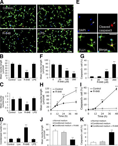
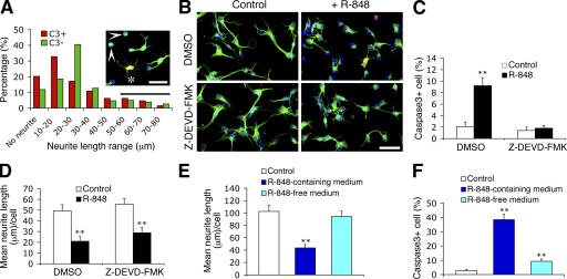
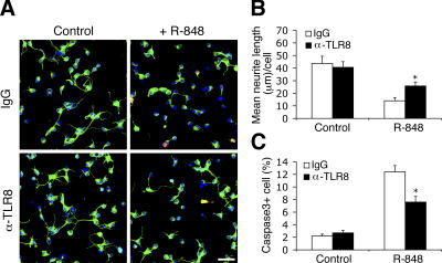
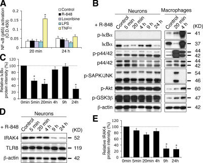
Toll receptors in Drosophila melanogaster function in morphogenesis and host defense. Mammalian orthologues of Toll, the Toll-like receptors (TLRs), have been studied extensively for their essential functions in controlling innate and adaptive immune responses. We report that TLR8 is dynamically expressed during mouse brain development and localizes to neurons and axons. Agonist stimulation of TLR8 in cultured cortical neurons causes inhibition of neurite outgrowth and induces apoptosis in a dissociable manner. Our evidence indicates that such TLR8-mediated neuronal responses do not involve the canonical TLR-NF-kappaB signaling pathway. These findings reveal novel functions for TLR8 in the mammalian nervous system that are distinct from the classical role of TLRs in immunity.


Figures

Figure 1.
Figure 1.
TLR8 is dynamically expressed during mouse brain development and localizes to axons and neurons. (A) Western blot analysis of TLR8 expression in the developing mouse brains. Spleen (Sp) and Raw264.7 (Raw) macrophages are positive controls for anti-TLR8 immunoreactivity. β-actin serves as loading control. (B) Immunohistochemical analysis of TLR8 expression in sagittal sections of E12 embryo, E18 brain, and P14 cerebral cortex. The images of the E12 embryo and E18 brain were acquired by confocal microscopy using the Tile Scan function. (C) Whole-mount in situ hybridization of E12 embryo using a digoxin (DIG)-labeled probe specific to Tlr8 mRNA. Arrowheads in B and C indicate the sympathetic nerve trunk. (D) Western blotting of TLR4, TLR7, TLR8, MyD88, and NF-κB subunit p65 in cortical neurons cultured for 1 (DIV1) and 5 d (DIV5). (E) Immunocytochemistry of TLR8 in cultured cortical neurons. MAP2 and neurofilament 200 kD (NFL) are neuron-specific markers. An affinity-purified anti-TLR8 polyclonal antibody was used in A, B, D, and E. P, postnatal day; P12w, 12-wk-old; ic, internal capsule; IMZ, cortical intermediate zone; f1, fimbria of hippocampus, OC, optic chiasm, ONL, olfactory nerve layer. Bars: (B, top, and C) 1 mm; (B, bottom) 100 μm; (E) 50 μm.
Figure 2.
Figure 2.
R-848 inhibits neurite outgrowth and triggers apoptosis in freshly cultured cortical neurons. (A) Representative micrographs of neurons untreated (control) or treated with 500 μM loxoribine (Lox; TLR7 agonist), 100 μM R-848 (TLR7/8 agonist), or 5 μg/ml LPS (TLR4 agonist) for 24 h. Cells were fixed and double-immunostained with anti–βIII-tubulin (green) and anti-cleaved caspase3 (red) monoclonal antibodies. (B–D) Quantification of the effects of PAMPs on neurite length (B), neurite number (C), and cell death (D) as characterized by condensed nuclei (<50 ± 25 μm2). (E) Representative micrographs of R-848–treated cultures show a neuron (βIII-tubulin/TUJ1) with a condensed nucleus (DAPI; arrowhead) stained by anti-cleaved caspase3. Dose–response curve (F and G) and time-course (H and I) of R-848 effects on neurite outgrowth (F and H) and apoptosis (G and I). In F and G, neurons were treated for 24 h; in H and I, neurons were exposed to 100 μM R-848. (J and K) Quantification of neurite length (J) and apoptosis (K) of neurons cultured for 24 h in normal or conditioned medium with or without 100 μM R-848. In a separate culture, neurons grown in normal medium were first stimulated with 100 μM R-848 for 18 h, washed thoroughly to eliminate trace amounts of R-848, and further incubated in fresh normal medium for 12 h, after which the supernatant was collected as the conditioned medium. Statistical analysis was done by t test. *, P < 0.05; **, P < 0.01 versus controls (untreated, 0 μM or 0 h). Data are the mean ± the SEM for triplicate determinations. Bars: (A) 100 μm; (E) 50 μm.
Figure 3.
Figure 3.
Neurite outgrowth inhibition and neuronal apoptosis induced by R-848 are dissociable. (A) Neurite length distribution of the cleaved caspase3-positive (C3+; n = 64) and the cleaved caspase3-negative neurons (C3−; n = 338) in cultures treated with 100 μM R-848 for 24 h. Cells with neurite length of <10 μm are defined as having no neurites, and cells with neurites longer than the average neurite length of the untreated neurons (56.4 ± 5.9 μm) are considered as having normal neurites (indicated by the horizontal bar). The insert shows a representative field of R-848–treated cultures. Asterisk and arrowheads indicate a C3+ cell with normal neurites and C3− cells with no neurites, respectively. Cells were fixed and double-immunostained with anti–βIII-tubulin (green) and anti-cleaved caspase3 (red) antibodies. (B) Representative micrographs of neurons untreated (control) or treated with 100 μM R-848 for 24 h in the presence of vehicle control (1% DMSO) or caspase3 inhibitor (20 μM Z-DEVD-FMK). (C and D) Quantification of the effects of blocking caspase3 activity on R-848–induced apoptosis (C) and neurite outgrowth inhibition (D). (E and F) Quantification of neurite length (E) and apoptosis (F) of neurons that were initially exposed to 100 μM R-848 for 12 h (the time when the R-848–induced morphological changes become detectable), then switched to R-848–free or –containing medium for a further incubation of 36 h. Control cultures were never exposed to R-848. Statistical analysis was done by t test. **, P < 0.01 versus controls. Data are the mean ± the SEM for triplicate determinations. Bars, 50 μm.
Figure 4.
Figure 4.
R-848 effects on neurons are attenuated by a polyclonal antibody specifically against TLR8. (A) Representative micrographs of nonimmune IgG- and anti-TLR8 (α-TLR8)–transduced neurons that were either untreated (control) or treated with 100 μM R-848 for 18 h. Cells were fixed and double-immunostained with anti–βIII-tubulin (green) and anti-cleaved caspase3 (red) antibodies. Bar, 50 μm. (B and C) Quantification of the anti-TLR8–mediated blocking effects on R-848–induced neurite outgrowth inhibition (B) and apoptosis (C). Statistical analysis was done by t test. *, P < 0.05 versus nonimmune IgG-transduced neurons treated with R-848. Data are the mean ± the SEM for triplicate determinations.
Figure 5.
Figure 5.
TLR8 stimulation in neurons does not activate the canonical TLR–NF-κB signaling pathway, but rather down-regulates IκBα and IRAK4. (A) ELISA assay for NF-κB (p65) transactivation using nuclear extracts from cortical neurons stimulated with 100 μM R-848, 500 μM loxoribine, 5 μg/ml LPS, or 10 ng/ml TNFα for the indicated times. LPS and TNFα serve as negative and positive controls, respectively. (B) Western blotting of the hallmarks of the conventional TLR-signaling pathway with lysates from neurons and Raw264.7 macrophages treated with 100 μM R-848 for the indicated times. (C) Quantification of changes in IκBα levels in R-848–stimulated neurons by band densitometry. A representative blot is shown in B. (D) Western blotting of IRAK4 in neurons stimulated with 100 μM R-848 for the indicated times. Note that TLR8 levels remain unchanged. (E) Quantification of changes in IRAK4 levels by band densitometry. A representative blot is shown in D. Data in C and E, expressed as percentage normalized to controls (100%), are the mean ± the SEM for pooled Western-blots from three independent cultures. Statistical analysis was done by t test. *, P < 0.05 versus controls.

References

    1. Akira, S., and K. Takeda. 2004. Toll-like receptor signalling. Nat. Rev. Immunol. 4:499–511. - PubMed
    1. Belvin, M.P., and K.V. Anderson. 1996. A conserved signaling pathway: the Drosophila toll-dorsal pathway. Annu. Rev. Cell Dev. Biol. 12:393–416. - PubMed
    1. Boulanger, L.M., and C.J. Shatz. 2004. Immune signalling in neural development, synaptic plasticity and disease. Nat. Rev. Neurosci. 5:521–531. - PubMed
    1. Coulpier, M., J. Anders, and C.F. Ibanez. 2002. Coordinated activation of autophosphorylation sites in the RET receptor tyrosine kinase: importance of tyrosine 1062 for GDNF mediated neuronal differentiation and survival. J. Biol. Chem. 277:1991–1999. - PubMed
    1. Du, X., A. Poltorak, Y. Wei, and B. Beutler. 2000. Three novel mammalian toll-like receptors: gene structure, expression, and evolution. Eur. Cytokine Netw. 11:362–371. - PubMed

Publication types

MeSH terms