. 2006 Oct 23;175(2):315-24.
doi: 10.1083/jcb.200603149.

Rapid actin monomer-insensitive depolymerization of Listeria actin comet tails by cofilin, coronin, and Aip1

Affiliations

Rapid actin monomer-insensitive depolymerization of Listeria actin comet tails by cofilin, coronin, and Aip1

William M Brieher et al. J Cell Biol. .

Abstract

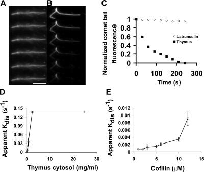
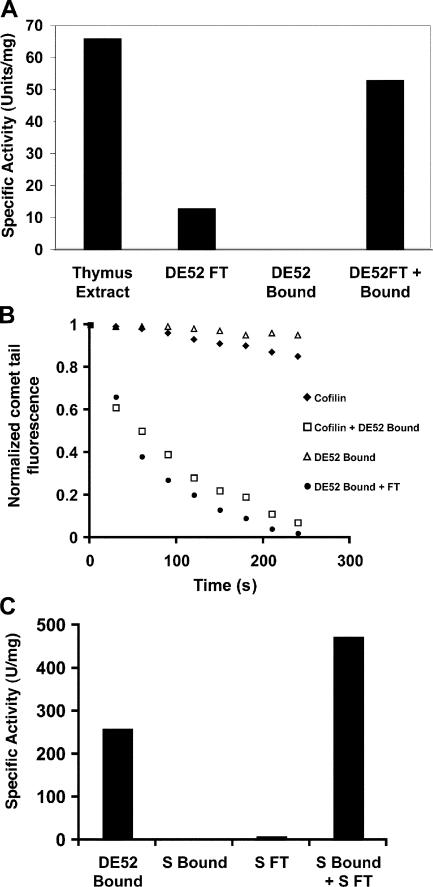
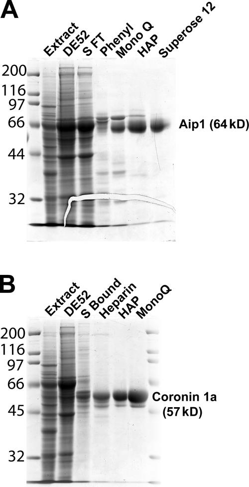
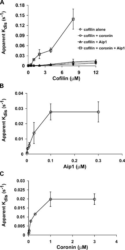
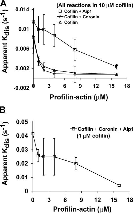
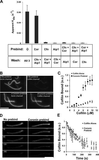
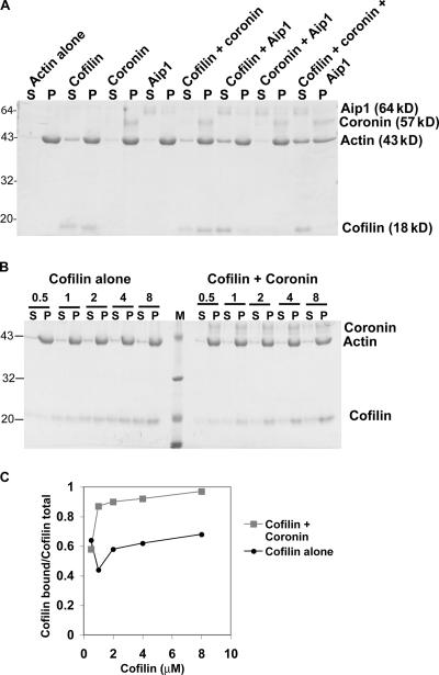
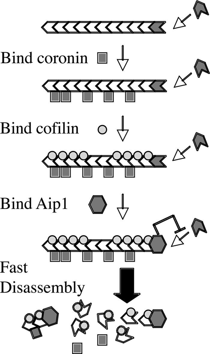
Actin filaments in cells depolymerize rapidly despite the presence of high concentrations of polymerizable G actin. Cofilin is recognized as a key regulator that promotes actin depolymerization. In this study, we show that although pure cofilin can disassemble Listeria monocytogenes actin comet tails, it cannot efficiently disassemble comet tails in the presence of polymerizable actin. Thymus extracts also rapidly disassemble comet tails, and this reaction is more efficient than pure cofilin when normalized to cofilin concentration. By biochemical fractionation, we identify Aip1 and coronin as two proteins present in thymus extract that facilitate the cofilin-mediated disassembly of Listeria comet tails. Together, coronin and Aip1 lower the amount of cofilin required to disassemble the comet tail and permit even low concentrations of cofilin to depolymerize actin in the presence of polymerizable G actin. The cooperative activities of cofilin, coronin, and Aip1 should provide a biochemical basis for understanding how actin filaments can grow in some places in the cell while shrinking in others.


Figures

Figure 1.
Figure 1.
Fast disassembly of Listeria actin comet tails requires cellular factors. (A) Frames from a time-lapse sequence showing the slow decay of comet tails diluted into latrunculin. (B) Time-lapse images of a Listeria comet tail in thymus extract. Frames in A and B are 30 s apart. (C) Actin intensity decay profiles of comet tails in latrunculin or thymus extract. Each point is the normalized mean of at least 30 comet tails from two experiments. (D and E) Apparent disassembly rates of comet tails in thymus extract (D) or pure cofilin alone (E). Mean ± SD (error bars); n = 3 experiments/point. Bar, 10 μm.
Figure 2.
Figure 2.
Thymus extract contains multiple factors that contribute to comet tail disassembly. (A) Thymus extract contains at least two factors involved in comet tail disassembly. One unit of activity is the amount of protein required to fully disassemble all comet tails in <5 min. (B) Cofilin substitutes for the thymus DE52 flow through fraction, whereas the DE52-bound fraction potentiates cofilin activity. (C) The DE52-bound cofilin-potentiating fraction consists of at least two factors. Specific activity is as described in A.
Figure 3.
Figure 3.
Purification of cofilin-potentiating factors from thymus extract. (A) Coomassie-stained gel showing the purification of factor X (Aip1). (B) Coomassie-stained gel showing the purification of factor Y (coronin-1A). Numbers left of the gels correspond to molecular mass standards in kilodaltons.
Figure 4.
Figure 4.
Dose-response analysis of cofilin, Aip1, and coronin in Listeria actin comet tail disassembly. (A) Amount of cofilin required to disassemble comet tails in the presence or absence of Aip1 and coronin. (B) Titration of the amount of Aip1 required to enhance the depolymerization of Listeria comet tails in the presence of 1 μM of limiting cofilin and 1 μM of saturating coronin. (C) Titration of the amount of coronin required to enhance the depolymerization of Listeria comet tails in the presence of 1 μM of limiting cofilin and 0.2 μM of saturating Aip1. Each plot shows the mean ± SD (error bars); n = 3 experiments/point.
Figure 5.
Figure 5.
Disassembly of Listeria actin comet tails in the presence of profilin–actin. (A) Comparison of comet tail disassembly rates as a function of profilin–actin concentration in the presence of a high concentration (10 μM) of cofilin alone versus 10 μM cofilin + 0.1 μM Aip1 or 10 μM cofilin + 1 μM coronin. (B) Comparison of comet tail disassembly rates as a function of profilin–actin concentration in the presence of all three depolymerizing factors. In this experiment, the cofilin was lowered to 1 μM while Aip1 was maintained at 0.1 μM and coronin was maintained at 1 μM. Each plot shows the mean ± SD (error bars); n = 3 experiments/point.
Figure 6.
Figure 6.
Coronin promotes cofilin binding to actin comet tails. (A) Apparent comet tail disassembly rates after the preincubation of comet tails in either buffer (φ), 1 μM coronin (Cor), 3 μM cofilin (Cfn), 0.3 μM Aip1, or a combination of any two factors. After preincubation, these solutions were replaced with the remaining factors at the same listed concentrations, and comet tail disassembly was measured by time-lapse imaging. Each experiment plots the mean ± SD (error bars); n = 4. (B) Paired fluorescent images of labeled cofilin bound to actin comet tails in the presence or absence of a coronin preincubation. (a and b) No preincubation. (c and d) Comet tails preincubated with 1 μM coronin. Both samples were then incubated in 4 μM of labeled cofilin before glutaraldehyde fixation and imaging. (a and c) Actin; (b and d) cofilin. (C) Ratio of cofilin fluorescence to actin fluorescence in the presence or absence of a coronin preincubation. n = 3 experiments for each point. (D) Images from a time-lapse sequence showing the dissociation of labeled cofilin from actin comet tails after a preincubation in buffer alone or cofilin from comet tails preincubated in 1 μM coronin. Numbers refer to time in seconds. (E) Comparison of cofilin dissociation rates from comet tails preincubated in buffer or 1 μM coronin. (C and D) Each point is the mean ± SD; n = 4 experiments. au, arbitrary units. Bars, 10 μm.
Figure 7.
Figure 7.
Sedimentation analysis of actin disassembly. (A) Coomassie gel of the distribution of combinations of actin, cofilin, coronin, and Aip1 between the supernatant (S) and pellet (P). (B) Coomassie gel of cofilin binding to F actin in the presence or absence of coronin. M, molecular mass markers. (C) Densitometry of gel shown in B. Numbers on the left of A and B are the molecular masses of standards in kilodaltons.
Figure 8.
Figure 8.
Model of actin filament disassembly by cofilin, coronin, and Aip1. Binding of coronin to a growing filament promotes cofilin binding, which, in turn, promotes binding of Aip1 to the barbed end. Aip1 inhibits elongation of the filament and triggers its fast disassembly. The binding of coronin, cofilin, and Aip1 contributes to a rate-limiting transition that converts a stable filament that can grow to an unstable filament, which rapidly shrinks.

References

    1. Abe, H., T. Obinata, L.S. Minamide, and J.R. Bamburg. 1996. Xenopus laevis actin-depolymerizing factor/cofilin: a phosphorylation-regulated protein essential for development. J. Cell Biol. 132:871–885. - PMC - PubMed
    1. Balcer, H.I., A.L. Goodman, A.A. Rodal, E. Smith, J. Kugler, J.E. Heuser, and B.L. Goode. 2003. Coordinated regulation of actin filament turnover by a high-molecular-weight Srv2/CAP complex, cofilin, profilin, and Aip1. Curr. Biol. 13:2159–2169. - PubMed
    1. Bamburg, J.R. 1999. Proteins of the ADF/cofilin family: essential regulators of actin dynamics. Annu. Rev. Cell Dev. Biol. 15:185–230. - PubMed
    1. Brieher, W.M., M. Coughlin, and T.J. Mitchison. 2004. Fascin-mediated propulsion of Listeria monocytogenes independent of frequent nucleation by the Arp2/3 complex. J. Cell Biol. 165:233–242. - PMC - PubMed
    1. Cameron, L.A., T.M. Svitkina, D. Vignjevic, J.A. Theriot, and G.G. Borisy. 2001. Dendritic organization of actin comet tails. Curr. Biol. 11:130–135. - PubMed

Publication types

MeSH terms