. 2006 Oct 31;103(44):16236-41.
doi: 10.1073/pnas.0604817103. Epub 2006 Oct 23.

Role of histone tails in chromatin folding revealed by a mesoscopic oligonucleosome model

Affiliations

Role of histone tails in chromatin folding revealed by a mesoscopic oligonucleosome model

Gaurav Arya et al. Proc Natl Acad Sci U S A. .

Abstract

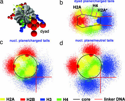
The role of each histone tail in regulating chromatin structure is elucidated by using a coarse-grained model of an oligonucleosome incorporating flexible histone tails that reproduces the conformational and dynamical properties of chromatin. Specifically, a tailored configurational-bias Monte Carlo method that efficiently samples the possible conformational states of oligonucleosomes yields positional distributions of histone tails around nucleosomes and illuminates the nature of tail/core/DNA interactions at various salt milieus. Analyses indicate that the H4 histone tails are most important in terms of mediating internucleosomal interactions, especially in highly compact chromatin with linker histones, followed by H3, H2A, and H2B tails in decreasing order of importance. In addition to mediating internucleosomal interactions, the H3 histone tails crucially screen the electrostatic repulsion between the entering/exiting DNA linkers. The H2A and H2B tails distribute themselves along the periphery of chromatin fibers and are important for mediating fiber/fiber interactions. A delicate balance between tail-mediated internucleosomal attraction and repulsion among linker DNAs allows the entering/exiting linker DNAs to align perpendicular to each other in linker-histone deficient chromatin, leading to the formation of an irregular zigzag-folded fiber with dominant pair-wise interactions between nucleosomes i and i +/- 4.


Conflict of interest statement

The authors declare no conflict of interest.

Figures

Fig. 1.
Fig. 1.
Modeling the basic motif c1 (nucleosome core, linker DNA, and histone tails) to yield the final model c2: the atomistic nucleosome core n1 is modeled as a rigid body with a uniformly distributed set of charges (n2); the linker DNA d1 is treated by using the discrete elastic chain model d2; and the histone tails [(t1), H3 tail is shown] are represented by using the subunit model t2 and then coarse-grained further to the protein bead chain t3. The tails in c2 are colored blue (H3), green (H4), yellow (H2A), and red (H2B).
Fig. 2.
Fig. 2.
Reference frame of a nucleosome core (a) and positional distribution of fully charged histone tails along its dyad (b) and nucleosomal planes (c) and of neutralized tails along a nucleosome plane (d), all at 0.2 M salt. Red arrows in b indicate the mean position/orientation of H4 and H2A tails. H2A*, C termini of H2A histones.
Fig. 3.
Fig. 3.
Representative 48-unit oligonucleosome at 0.2 M salt (a) and analyses and snapshots of boxed regions in a to highlight different tail interactions (b–g%). Analyzed systems involve oligonucleosomes without linker histones at 0.2 M (□) and 0.01 M (▵), and “compact” oligonucleosomes at 0.2 M salt (×). In each box (b–g%), a cartoon image depicts the interaction plotted as a frequency for the time that tails mediate: internucleosomal interactions (b), attach to parent nucleosomes (c), remain unattached (d), attach to linker DNA not associated with parent nucleosome (e), and attach to linker DNA associated with parent nucleosome (f). Plot (g) provides tail extension lengths. Results are averaged over the two copies of each tail. H2A*, C termini of H2A histones.
Fig. 4.
Fig. 4.
Representative configurations of oligonucleosomes with 6 (a), 12 (b), 24 (c), and 48 (d) nucleosomes at 0.2 M salt. The nucleosome cores are shown as white cylinders, and the DNAs are shown as red cylindrical tubes. The histone tails are omitted for clarity.
Fig. 5.
Fig. 5.
Internucleosomal interactions within a 48-unit oligonucleosome at 0.2 M salt. Color-coded map showing intensity of interaction between nucleosome i and j (a) and interaction intensity I(k) versus linker DNA separation k (b).

References

    1. Felsenfeld G, Groudine M. Nature. 2003;421:448–453. - PubMed
    1. Richmond TJ, Davey CA. Nature. 2003;423:145–150. - PubMed
    1. Tse C, Sera T, Wolffe AP, Hansen JC. Mol Cell Biol. 1998;18:4629–4638. - PMC - PubMed
    1. Wade PA, Pruss D, Wolffe AP. Trends Bio Sci. 1997;22:128–132. - PubMed
    1. Davie JR. In: Chromatin Structure and Dynamics: State-of-the-Art. Zlatanova J, Leuba SH, editors. Amsterdam, The Netherlands: Elsevier; 2004. pp. 205–240.

Publication types

LinkOut - more resources