. 1991 Mar 7;350(6313):68-71.
doi: 10.1038/350068a0.

Cell-surface regulation of beta 1-integrin activity on developing retinal neurons

Affiliations

Cell-surface regulation of beta 1-integrin activity on developing retinal neurons

K M Neugebauer et al. Nature. .

Abstract

Integrins are a family of alpha beta heterodimeric receptors that mediate cell-cell and cell-substratum interactions. Integrin binding to extracellular ligands regulates cell adhesion, shape, motility, intracellular signalling and gene expression. Mechanisms that regulate integrin function are, therefore, central to the participation of integrins in a diverse set of cellular events. Here we report the identification of TASC, a monoclonal antibody to a novel epitope on the integrin beta 1 subunit, which inhibits cell adhesion to vitronectin but promotes adhesion to laminin and collagen types I and IV. We show that developing retinal neurons that have lost responsiveness to laminin regain the ability to bind laminin in the presence of TASC. Thus, beta 1-class integrins are likely to occupy multiple affinity states that can be modulated at the cell surface.


Figures

FIG. 1
FIG. 1
TASC inhibits E7 retinal neuron attachment to vitronectin and promotes attachment to laminin and collagens. a, Attachment to vitronectin is inhibited by l00 μg m−1 GRGDSP and 200 μg ml−1 TASC, 50 μg ml−1 TASC Fab, and 50 μg ml−1 CSAT. b, Histograms of retinal neuron attachment to collagen IV (10 min), laminin (20 min) and fibrinogen (1 h). All antibodies are used at 50 μg ml−1. c, Time-course of E7 retinal neuron attachment to collagen I in the presence of no antibody, TASC IgG, TASC Fab and TASC IgG plus CSAT. d, Attachment of E7 retinal neurons to a collagen-I concentration curve at 40 min in the presence of no antibody or TASC at 25 μg ml−1. All values are expressed as a percentage of the attachment observed on poly-d-lysine which is considered to represent a signal of 100%. METHODS. Vitronectin was purified from fetal bovine serum by chromatography on Heparin Sepharose Cl-6b (Pharmacia). Collagen I (Collaborative Research) was diluted to 10 μg ml−1 in 0.1 N acetic acid. Collagen IV (Collaborative Research) was diluted to 10 μg ml−1 in CMF-PBS. Laminin (diluted to 20 (μg ml−1 in CMF-PBS) was purified from murine EHS tumour by I. de Curtis and M. J. Ignatius according to published procedures. Fibrinogen was the gift of Z. Ruggeri (Research Institute of the Scripps Clinic, La Jolla, California) and was used at 20 μg ml−1 in Ca2+/Mg2+ containing PBS. Poly-d-lysine was from Sigma. IgGs were purified from ascites fluid on protein A/Sepharose Cl-4b (Pharmacia), and GRGDSP was purchased from Collaborative Research. TASC Fab fragments were generated from purified IgG by papain (Sigma) digestion followed by ion-exchange chromatography on DE-52 (What-man). Cell attachment was quantitated as previously described, except that adherent cells were stained with crystal violet and adsorbtion at 540 nm was subsequently measured. In each experiment, attachment to BSA-coated wells was subtracted, and all determinations were done in triplicate. To isolate TASC, integrin-rich chick brain glial cells were use d as the immunogen for monoclonal antibody production in mice. Clonal supernatants were screened for the ability to inhibit retinal neuron attachment to vitronectin.
FIG. 2
FIG. 2
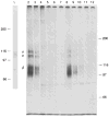
TASC recognizes the integrin β1 subunit. TASC binds a 105-110K band in an immunoblot of E7 retinal extracts (lane 1). Both TASC and CSAT immunoprecipitate the integrin β1 subunit of 100K and 110K and associated α subunits at 135K and 145K. Immunodepletion analysis of the TASC antigen on E7 retinal neurons reveals that sequential precipitation of labelled extract with CSAT (lanes 2–5) removes the TASC antigen (lane 6). Overexposure of the autoradiogram did not reveal the presence of even minor amounts of either antigen following depletion (not shown). Antigens were unlikely to have been degraded during the course of the experiment, because another cell-surface glycoprotein, the integrin αv subunit, was readily detected by immunoprecipitation from an aliquot of the same depleted sample (lane 7). Depletion with TASC (lanes 8–11) removes the CSAT antigen (lane 12). METHODS. E7 retinas were extracted for 15 min in an equal volume of ice-cold Ca2+/Mg2+-containing PBS plus 1% Triton X-100 and 2 mM PMSF. Supernatant (200 μg) was run under nonreducing conditions on a 6% gel and transferred to nitrocellulose. The blots were incubated with 20 μg ml−1 TASC overnight at 4 °C, followed by alkaline phosphatase-conjugated second antibody (Promega), used and developed according to manufacturer’s instructions. For immunoprecipitation, E7 retinal neurons were metabolically labelled with 100 μCi ml−1 [35S] methionine/cysteine (NEN) for 16 h. Cells were collected and extracted as above. The supernatant was divided into two 200 μl aliquots, and 20 μg TASC or CSAT was added to each. Samples were rocked overnight at 4 °C and then precleared twice with 50 μl of Sepharose Cl-4b (Pharmacia). Antibody-antigen complexes were precipitated with protein A/Sepharose CI-4B and washed extensively with 0.05M Tris buffer pH 8.0, 150 mM NaCl, 1 mM CaCl2, 1 mM MgCl2, 0.5% Triton X-100. To deplete the extract of antigen, supernatants were repeatedly precipitated with 20 μg of each antibody. An additional protein A/Sepharose adsorption step was performed before adding the second antibody to ensure that all of the depleting antibody had been removed. The resulting supernatant was divided in half and precipitated with 20 μg of the second antibody. The monoclonal antibody to αv was isolated in this laboratory. Pellets were extracted in sample buffer and separated by nonreducing SDS-PAGE on 6% gels. Gels were fixed, stained, treated with En3Hance (NEN), dried, and exposed to Kodak X Omat R film. Mr standards were obtained from Biorad.
FIG. 3
FIG. 3
E11 retinal neurons do not adhere to laminin in the absence of TASC at any time point up to 60 min (open squares). In the presence of 100 μg ml−1 TASC (●), attachment to laminin is detectable within 10 min and peaks at 40 min with ~80 % of the neurons attached compared to poly-d-lysine. TASC-induced attachment is completely blocked by the addition of CSAT (■).

References

    1. Akiyama SK, Nagata K, Yamada KM. Biochim biophys Acta. 1990;1031:91–110. - PubMed
    1. Hemler ME. A Rev Immun. 1990;8:365–400. - PubMed
    1. Springer TA. Nature. 1990;346:425–434. - PubMed
    1. Hall DE, Neugebauer KM, Reichardt LF. J Cell Biol. 1987;104:623–634. - PMC - PubMed
    1. Reichardt LF, Tomaselli KJ. A Rev Neurosci. 1991;14:531–570. - PMC - PubMed

Publication types

MeSH terms