. 2006 Dec;116(12):3229-39.
doi: 10.1172/JCI29867. Epub 2006 Oct 19.

Ghrelin modulates the activity and synaptic input organization of midbrain dopamine neurons while promoting appetite

Affiliations

Ghrelin modulates the activity and synaptic input organization of midbrain dopamine neurons while promoting appetite

Alfonso Abizaid et al. J Clin Invest. 2006 Dec.

Abstract

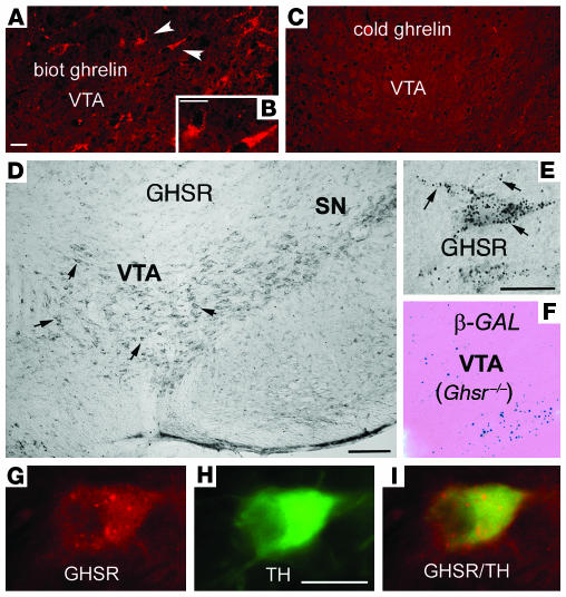
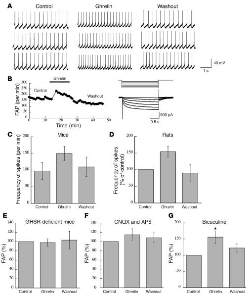
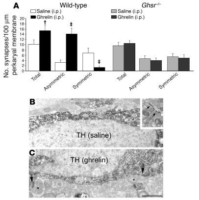
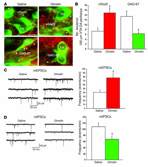
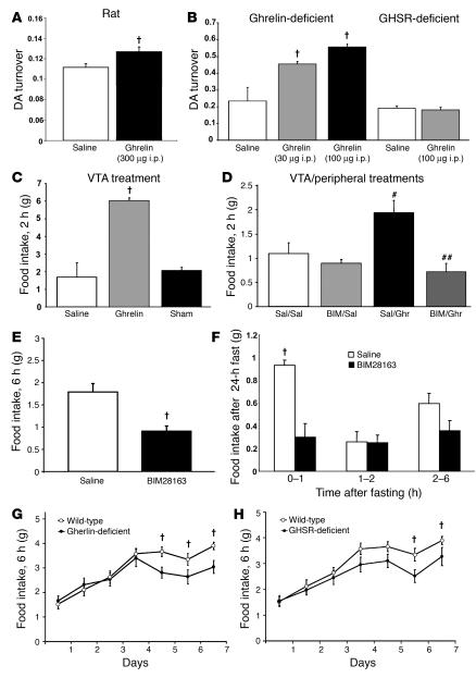
The gut hormone ghrelin targets the brain to promote food intake and adiposity. The ghrelin receptor growth hormone secretagogue 1 receptor (GHSR) is present in hypothalamic centers controlling energy metabolism as well as in the ventral tegmental area (VTA), a region important for motivational aspects of multiple behaviors, including feeding. Here we show that in mice and rats, ghrelin bound to neurons of the VTA, where it triggered increased dopamine neuronal activity, synapse formation, and dopamine turnover in the nucleus accumbens in a GHSR-dependent manner. Direct VTA administration of ghrelin also triggered feeding, while intra-VTA delivery of a selective GHSR antagonist blocked the orexigenic effect of circulating ghrelin and blunted rebound feeding following fasting. In addition, ghrelin- and GHSR-deficient mice showed attenuated feeding responses to restricted feeding schedules. Taken together, these data suggest that the mesolimbic reward circuitry is targeted by peripheral ghrelin to influence physiological mechanisms related to feeding.


Figures

Figure 1
Figure 1. Expression of GHSR and ghrelin binding in VTA neurons of the rat.
(AC) Rat VTA sections incubated in either biotinylated (biot) ghrelin (arrowheads; A and B) or unlabeled (cold) ghrelin (C). (D and E) GHSR immunocytochemistry in mouse (D) and rat (E) brain sections. Dense immunolabeling was observed in the VTA (D, arrows), with cells showing bouton-like immunoreactivity (IR; E, arrows). SN, substantia nigra. (F) β-Gal–labeled blue nuclei in the VTA of Ghsr–/– mice confirmed expression of GHSR. (GI) High-magnification images of a GHSR-immunolabeled (red) cell that coexpressed TH (green). Scale bars: 10 μm (AC, E, and GI); 100 μm (D and F).
Figure 2
Figure 2. Ghrelin increases action potential generation in VTA DA neurons in mice (n = 9) and rats (n = 6).
(A) Raw traces recorded before (Control), during (Ghrelin), and after (Washout) application of ghrelin. (B) Time course of the ghrelin-induced increase in frequency of action potential (FAP). AC represent time points when traces from A were recorded. (C and D) Mean frequency of action potentials recorded in mice (C) and rats (D) before, during, and after application of ghrelin. Hyperpolarization-induced action current recorded from DA neurons is shown (D, inset). (E and F) Ghrelin-mediated enhancement of action potential generation in VTA DA neurons required excitatory inputs. Shown are mean frequency of action potentials recorded before, during, and after application of ghrelin. (E) No effect of ghrelin was observed on frequency of action potentials in slices from GHSR-knockout mice (n = 5). (F) In the absence of excitatory input onto DA neurons, no effect of ghrelin was observed on frequency of action potentials in slices from wild-type mice (n = 6) in the presence of CNQX (10 μM) and AP5 (50 μM) in all solutions. Results were pooled from all recorded neurons. (G) Experiments were performed in slices from wild-type mice (n = 7) in the presence of bicuculline (30 μM) in all solutions. Results were pooled from all recorded neurons. In the absence of inhibitory inputs of DA neurons, ghrelin elevated the frequency of action potentials of VTA DA neurons, an effect that diminished after washout.
Figure 3
Figure 3. Synaptic remodeling induced by peripheral ghrelin in VTA DA cells.
(A) Ghrelin increased the number of synapses on VTA DA cells of wild-type mice (n = 5). Both total and asymmetric synaptic contacts were elevated, while the number of symmetric synapses was decreased. No synaptic changes were observed after peripheral ghrelin injection in Ghsr–/– mice (n = 5). P < 0.05, P < 0.01 versus respective saline-treated controls. (B and C) Electron micrographs showing typical TH-immunoreactive perikarya of the VTA from saline- (B) and ghrelin-treated (C) wild-type mice. Arrows in inset of B indicate a symmetric synapse. Arrows in C indicate asymmetric synaptic contacts. Asterisks indicate unlabeled axon terminals. Scale bar: 1 μm.
Figure 4
Figure 4. Ghrelin alters inhibitory and excitatory inputs of VTA DA cells.
(A and B) Appositions between vGlut2-immunoreactive (red) and TH-immunopositive (green) VTA perikarya were significantly greater in ghrelin-treated animals compared with saline-treated controls. In contrast, appositions between GAD-67–immunolabeled boutons and TH-immunoreactive VTA perikarya were significantly lower in ghrelin-treated animals compared with saline-treated controls. Scale bars: 10 μm. (C) Corresponding to the observed changes in the number of synapses by light and electron microscopy, ghrelin treatment induced a significant elevation in the frequency of mEPSCs compared with saline controls. (D) Conversely, ghrelin administration triggered a significant decrease in the frequency of mIPSCs that was also in line with the light and electron microscopy results. P < 0.05 versus respective saline-treated controls.
Figure 5
Figure 5. Ghrelin alters dopamine turnover and feeding via the VTA.
(A) Peripheral ghrelin treatment (1 mg/kg) was effective in increasing DA turnover in the nucleus accumbens of rats (n = 12). (B) Ghrelin treatment (30 or 100 μg) of ghrelin-deficient mice increased DA turnover in the ventral striatum in a dose-dependent manner compared with saline-treated mice. Conversely, ghrelin induced no alterations in DA turnover of the nucleus accumbens in Ghsr–/– mice (n = 5 per treatment). (C) VTA ghrelin infusions (0.5 μg in 0.5 μl saline) significantly increased food intake in rats compared with saline infusions or with infusions at sites adjacent to, but not in, the VTA (Sham). (D) Ghrelin (5 μg in 0.1 ml saline) injected i.p. increased food intake compared with saline-injected rats, an effect blocked by BIM28163 infusion (0.5 ng in 0.5 μl saline) directly into the VTA. (E and F) Rebound feeding 6 hours after fasting was significantly attenuated in mice treated with BIM12863 (1 nM/d) infused into the VTA at 0.25 μl/h (E). This effect was statistically significant only during the first hour after food was reintroduced (F). (G and H) Six-hour food intake in ghrelin- (G) and GHSR-deficient (H) mice under a restricted feeding schedule. Both ghrelin- and GHSR-deficient mice showed attenuated feeding responses after repeated overnight fasts. P < 0.05 versus respective controls. #P < 0.05 versus saline/saline and BIM28163/saline treatment groups; ##P < 0.05 versus saline/ghrelin treatment group.

References

    1. Van der Lely A.J., Tschop M., Heiman M.L., Ghigo E. Biological, physiological, pathophysiological, and pharmacological aspects of ghrelin. Endocr. Rev. 2004;25:426–457. - PubMed
    1. Kojima M., et al. Ghrelin is a growth-hormone-releasing acylated peptide from stomach. Nature. 1999;402:656–660. - PubMed
    1. Tschop M., Smiley D.L., Heiman M.L. Ghrelin induces adiposity in rodents. Nature. 2000;407:908–913. - PubMed
    1. Nakazato M., et al. A role for ghrelin in the central regulation of feeding. Nature. 2001;409:194–198. - PubMed
    1. Cowley M.A., et al. The distribution and mechanism of action of ghrelin in the CNS demonstrates a novel hypothalamic circuit regulating energy homeostasis. Neuron. 2003;37:649–661. - PubMed

Publication types

MeSH terms