

Keystones in lymph node development
- PMID: 17062017
- PMCID: PMC2100342
- DOI: 10.1111/j.1469-7580.2006.00650.x
Keystones in lymph node development
Abstract
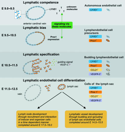
New molecular markers are constantly increasing our knowledge of developmental processes. In this review article we have attempted to summarize the keystones of lymphoid tissue development in embryonic and pathological conditions. During embryonic lymph node development in the mouse, cells from the anterior cardinal vein start to bud and sprout, forming a lymph sac at defined sites. The protrusion of mesenchymal tissue into the lymph sacs forms the environment, where so-called 'lymphoid tissue inducer cells' and 'mesenchymal organizer cells' meet and interact. Defects of molecules involved in the recruitment and signalling cascades of these cells lead to primary immunodeficiency diseases. A comparison of molecules involved in the development of secondary lymphoid organs and tertiary lymphoid organs, e.g. in autoimmune diseases, shows that the same molecules are involved in both processes. This has led to the hypothesis that the development of tertiary lymphoid organs is a recapitulation of embryonic lymphoid tissue development at ectopic sites.
Figures
References
-
- Adachi S, Yoshida H, Honda K, et al. Essential role of IL-7 receptor α in the formation of Peyer’s patch anlage. Int Immunol. 1998;10:1–6. - PubMed
-
- Aloisi F, Pujol-Borrell R. Lymphoid neogenesis in chronic inflammatory diseases. Nat Rev Immunol. 2006;6:205–217. - PubMed
-
- Ansel KM, Ngo VN, Hyman PL, et al. A chemokine-driven positive feedback loop organizes lymphoid follicles. Nature. 2000;406:309–314. - PubMed
-
- Ansel KM, Cyster JG. Chemokines in lymphopoiesis and lymphoid organ development. Curr Opin Immunol. 2001;13:172–179. - PubMed
Publication types
MeSH terms
Substances
LinkOut - more resources
Full Text Sources
