. 2006 Nov;209(5):611-22.
doi: 10.1111/j.1469-7580.2006.00646.x.

Micro-anatomical response of cartilage-on-bone to compression: mechanisms of deformation within and beyond the directly loaded matrix

Affiliations

Micro-anatomical response of cartilage-on-bone to compression: mechanisms of deformation within and beyond the directly loaded matrix

Ashvin Thambyah et al. J Anat. 2006 Nov.

Abstract

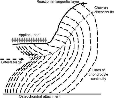
The biomechanical function of articular cartilage relies crucially on its integration with both the subchondral bone and the wider continuum of cartilage beyond the directly loaded contact region. This study was aimed at visualizing, at the microanatomical level, the deformation response of cartilage including that of the non-directly loaded continuum. Cartilage-on-bone samples from bovine patellae were loaded in static compression until a near-equilibrium deformation was achieved, and then chemically fixed in this deformed state. Full-depth cartilage-bone sections, incorporating the indentation profile and beyond, were studied in their fully hydrated state using differential interference contrast microscopy. Morphometric measurements of the indented profile were used in combination with a force analysis of the tangential layer to investigate the extent to which the applied force is attenuated in moving away from the directly loaded region. This study provides microscopic evidence of a structure-related response in the transitional zone of the cartilage matrix. It is manifested as an intense chevron-type shear discontinuity arising from the constraints provided by both the strain-limiting articular surface and the osteochondral attachment. The discontinuity persists well into the non-directly loaded continuum of cartilage and is proposed as a force attenuation mechanism. The structural and biomechanical analyses presented in this study emphasize the important role of the complex microanatomy of cartilage, highlighting the interconnectivity and optimal recruitment of the load-bearing elements throughout the zonally differentiated cartilage depth.


Figures

Fig. 1
Fig. 1
(A) Schematic of a typical diametrical section showing L0 and L, lengths before and after compression, respectively, determined with the aid of the chondrocytes, which acted as markers of the overall deformation field in the underlying matrix. Strain ɛ is calculated from these lengths. (B) Schematic of the force diagram based on a simplified geometry of indentation at equilibrium.
Fig. 2
Fig. 2
A typical diametrical section of a compressed osteochondral sample in its postfixed state. Scale bar = 2 mm.
Fig. 3
Fig. 3
(A) Typical pattern of deformation in the axi-symmetric region of loading (see dotted vertical line) under DIC imaging. AS = articular surface; TM = tidemark. Scale bar = 200 µm. (B) Deformation field extending laterally into the edge effect region and beyond (DIC). Scale bar = 200 µm. (C) Higher magnification view of edge effect region and beyond showing a diminishing intensity of the chevron discontinuity in the non-directly loaded cartilage continuum. Dashed line indicates the approximate position of the transition zone in the region where the discontinuity has almost vanished. Scale bar = 100 µm. (D) The development of tensile ‘pull-lines’ can be seen in the tangential zone beyond the directly loaded region. Scale bar = 200 µm.
Fig. 4
Fig. 4
Higher resolution DIC image of the chevron discontinuity (marked CD) in the transition zone. The articular surface is indicated (AS). Scale bar = 100 µm.
Fig. 5
Fig. 5
Higher magnification views of the transition from the mid-zone (MZ) towards the articular surface (AS) showing in image (A) a strong radial texture in the mid-zone; in image (B) a much less pronounced directional texture. Note that in both images there is a strong refractile boundary (RB) effect in the transition region. Scale bar = 50 µm.
Fig. 6
Fig. 6
Higher magnification view of the osteochondral junction: (A) directly loaded region in which the chondrocytes indicate matrix shear right down to the tidemark (TM), and (B) the non-loaded region for comparison. Scale bar = 100 µm.
Fig. 7
Fig. 7
(A) Schematic illustrating the ‘lines of chondrocyte continuity’ in relation to collagen fibril alignment and interconnectivity. This figure conveys the relative homogeneity of the collagenous architecture with the chondrocytes delineating the overall direction of fibrillar orientation. (B) The lines of chondrocyte continuity provide an ‘imprint’ of the overall arrangement of fibrils throughout the various zones.
Fig. 8
Fig. 8
Schematic tracing the chondrocyte continuity lines near the edge-effect region. There is a progressive dissipation of force, as indicated by the chevron discontinuity changing from an acute rectilinear form to curvilinear with increasing distance into the non-directly loaded region.

References

    1. Akizuki S, Mow VC, Muller F, Pita JC, Howell DS, Manicourt DH. Tensile properties of human knee joint cartilage. I. Influence of ionic conditions, weight bearing, and fibrillation on the tensile modulus. J Orthop Res. 1986;4:379–392. - PubMed
    1. Benninghoff A. Form und bau der Gelenknorpel in ihren Bezeihungen zur Funktion. Z Zellforsch Mikrosk Anat. 1925;2:783–825.
    1. Broom ND. Abnormal softening in articular cartilage: its relationship to the collagen framework. Arthritis Rheum. 1982;25:1209–1216. - PubMed
    1. Broom ND. Further insights into the structural principles governing the function of articular cartilage. J Anat. 1984;139:275–294. - PMC - PubMed
    1. Broom ND, Marra DL. New structural concepts of articular cartilage demonstrated with a physical model. Connect Tissue Res. 1985;14:1–8. - PubMed

Publication types

LinkOut - more resources