

Craniofacial levels and the morphological maturation of the human skull
- PMID: 17062021
- PMCID: PMC2100348
- DOI: 10.1111/j.1469-7580.2006.00644.x
Craniofacial levels and the morphological maturation of the human skull
Abstract
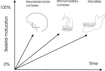
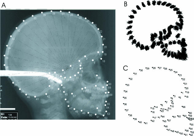
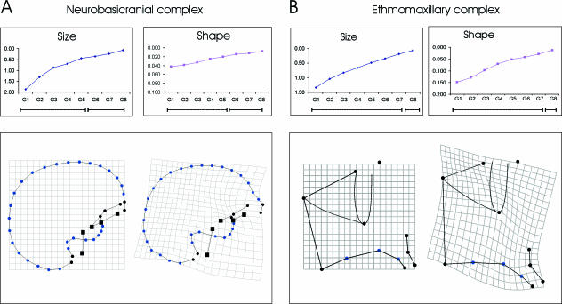
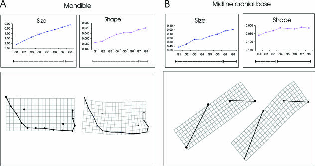
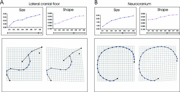
It is well known that the human skull achieves adult size through a superior-inferior gradient of maturation. Because the basicranium matures in size before the face, it has been suggested that the form of the basicranium might have ontogenetic knock-on effects on that of the face. However, although sequential spatially organized maturation of size is well described in the cranium, the maturation of skull shape is not. Knowledge of the maturation of shape is important, nevertheless, because it is claimed that the early determination of the spatial configuration of basicranial components, where the facial skeleton attaches, is relevant in the spatio-temporal ontogenetic cascade from basicranium to face. This paper examines the ontogeny of various components of the human skull in 28 individuals from the longitudinal Denver Growth Study. Sixty-six landmarks and semilandmarks were digitized on 228 X-rays and analysed using geometric morphometric methods. Bootstrapped confidence intervals for centroid size support previous studies suggesting a supero-inferior gradient of growth maturation (size over time), while developmental maturation (shape over time) is more complex. A sequence of shape maturation is described, in which the earliest structure to mature in shape was the midline cranial base (7-8 years), followed by the lateral cranial floor (11-12), midline neurocranium (9-10) and facial and mandibular structures (15-16). The absolute ages of shape maturation of the latter three depended on the criterion of maturity used, which was not the case for the basicranial components. Additionally, ontogenetic dissociations were found between the maturation of size and shape of the midline cranial base and lateral floor, possibly underlining its role as structural 'interface' between brain and facial ontogeny. These findings imply potential for bidirectional developmental influences between the lateral cranial floor and the face until about 11-12 years. The findings are discussed with regard to their relevance for palaeoanthropology and especially the evolutionary and developmental bases of skull morphological variation.
Figures
References
-
- Atchley WR, Hall BK. A model for development and evolution of complex morphological structures. Biol Rev. 1991;66:101–157. - PubMed
-
- Baba H, Aziz F, Kaifu Y, Suwa G, Kono RT, Jacob T. Homo erectus Calvarium from the Pleistocene of Java. Science. 2003;299:1384–1388. - PubMed
-
- Bastir M, Rosas A, Kuroe K. Morphogenetic determinants of mandibular ramus breadth. A test in modern human populations. Am J Phys Anthropol. 2002;S115:40–41.
-
- Bastir M. PhD thesis. Department of Anthropology, Autonoma University of Madrid; 2004. A geometric morphometric analysis of integrative morphology and variation in human skulls with implications for the Atapuerca-SH hominids and the evolution of Neandertals. Structural and systemic factors of morphology in the hominid craniofacial system.
-
- Bastir M, Rosas A. Comparative ontogeny in humans and chimpanzees: Similarities, differences and paradoxes in postnatal growth and development of the skull. Ann Anat. 2004a;186:503–509. - PubMed
Publication types
MeSH terms
LinkOut - more resources
Full Text Sources
Medical
