. 2006 Nov;209(5):655-69.
doi: 10.1111/j.1469-7580.2006.00643.x.

Analysis of the surface characteristics and mineralization status of feline teeth using scanning electron microscopy

Affiliations

Analysis of the surface characteristics and mineralization status of feline teeth using scanning electron microscopy

A DeLaurier et al. J Anat. 2006 Nov.

Abstract

External resorption of teeth by odontoclasts is a common condition of unknown origin affecting domestic cats. Odontoclastic resorptive lesions involve the enamel cementum junction (ECJ, cervix) and root surface, leading to extensive loss of enamel, dentine and cementum. This study was undertaken in order to determine whether features of the surface anatomy and mineralization of feline teeth could explain why odontoclastic resorptive lesions are so prevalent in this species. Backscattered electron scanning electron microscopy was used to study enamel, cementum and dentine in non-resorbed, undemineralized teeth from adult cats. Analysis of the ECJ revealed thin enamel and cementum and exposed dentine at this site. Furthermore, enamel mineralization decreased from the crown tip to the ECJ, and dentine mineralization was lowest at the ECJ and cervical root. Analysis of cementum revealed variations in the organization and composition of fibres between the cervical, mid- and apical root although no significant differences in mineralization of cementum were detected between different regions of the root. Reparative patches associated with resorption of cementum by odontoclasts and repair by cementoblasts were present on the root surface. In conclusion, results suggest that the ECJ and cervical dentine could be at a greater risk of destruction by odontoclasts compared with other regions of the tooth. The relationship of these features to the development and progression of resorption now requires further examination.


Figures
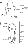
Fig. 1
Fig. 1
Schematic diagrams illustrating the sectioning and analysis of methacrylate-embedded teeth. (A) Teeth were sectioned longitudinally, slightly offset from the centre of roots (plane of section indicated by dotted lines). Both the mesial and the distal roots of multirooted premolars and molars were sectioned and analysed separately. The specimen surface was ground until the surface was as close to the central plane of the root as possible. (B) Locations where mineralization was measured in sectioned teeth. 1. Crown tip enamel (surface enamel, inner enamel, and enamel dentine junction) and crown tip dentine. 2. Mid-crown enamel (surface enamel and enamel dentine junction) and mid-crown dentine. 3. Enamel and dentine and the enamel cementum junction. 4. Cervical root cementum and dentine. 5. Mid-root cementum (surface cementum and cementum dentine junction) and dentine. 6. Apical root cementum (surface cementum and cementum dentine junction) and dentine.
Fig. 2
Fig. 2
Surface and section features of enamel (EP = enamel prisms). (A) Enamel prisms of a left upper first molar exposed through wear (scale bar = 25 µm). (B) Developing enamel surface morphology of a lower right fourth premolar showing enamel prisms (pit floor) and interpit enamel (IP, scale bar = 20 µm). (C) Developing enamel surface morphology showing ‘cobbles’ (CB) surrounded by prism-free enamel on the crown surface coronal to the enamel cementum junction (ECJ) in a left upper fourth premolar (scale bar = 25 µm). (D) Enamel of a lower left third premolar fractured post-mortem, showing horizontal sections through enamel prisms, which are orientated in alternate directions forming Hunter–Schreger bands, the prism-free surface enamel layer (PFE), and the enamel dentine junction (EDJ) (scale bar = 50 µm).
Fig. 3
Fig. 3
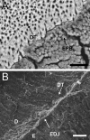
Surface and section features of dentine. (A) Dentine tubules (DT) exposed at the cervical root surface adjacent to cementum featuring extrinsic fibre bundles (EFB) on a left upper third premolar. (B) Upper left third premolar fractured post-mortem, showing dentine tubules (DT) radiating from the pulp chamber to the enamel dentine junction (EDJ) (D = dentine, E = enamel). All scale bars = 25 µm.
Fig. 4
Fig. 4
Surface features of the enamel cementum junction (ECJ) (E = enamel, RS = root surface). (A) ‘Cobbled’ (CB) enamel at the ECJ associated with a root surface featuring cementum with extrinsic fibre bundles (EFB) on a lower left third premolar (scale bar = 25 µm). (B) Enamel margin (EM) with enamel tubules (ET) and exposed dentine tubules (DT) on the root surface on a lower right canine (scale bar = 25 µm). (C) Cementum (CE) overlapping enamel on the root surface of an upper right fourth premolar (scale bar = 50 µm).
Fig. 5
Fig. 5
Features of extrinsic and intrinsic fibre cementum, and cementocyte lacunae on root surfaces (EFB = extrinsic fibre bundles, IF = intrinsic fibres, CL = cementocyte lacunae). (A) Intrinsic fibre cementum distributed between projecting extrinsic fibre bundles on a left lower third premolar (scale bar = 25 µm). (B) Cementum surface of a left lower third premolar fractured post-mortem showing extrinsic fibre bundles that can be traced from the cementum dentine junction (CDJ) to the root surface (RS) (scale bar = 25 µm). (C) Patch of remodelling intrinsic fibre cementum on the root surface of an upper right fourth premolar. Cementocyte lacunae are present among intrinsic fibres (scale bar = 25 µm). (D) Cementocyte lacunae among extrinsic and intrinsic fibres on an upper right first molar. Canaliculi can be observed in the base of lacunae (white arrow, scale bar = 10 µm).
Fig. 6
Fig. 6
Features of enamel in sectioned teeth (EP = enamel prisms, IP = interpit ameloblastic enamel, EDJ = enamel dentine junction). (A) Enamel prisms surrounded by interpit enamel at the EDJ of a lower right canine. Enamel prisms are organized in alternating Hunter–Schreger bands (scale bar = 50 µm). (B) Enamel tubules (ET) at the EDJ of a lower right first molar (scale bar = 50 µm). (C) Prism-free enamel (PFE) at the crown surface of a lower right canine (scale bar = 20 µm).
Fig. 7
Fig. 7
Features of the enamel cementum junction (ECJ) structure in sectioned teeth (CE = cementum, E = enamel). (A) Edge-to-edge contact between enamel and cementum in a lower left third premolar. (B) Enamel extending as a thin projection to meet cementum in a lower left third premolar. (C) Enamel margin (EM) joining exposed dentine (D) in a lower left canine. Scale bars = 50 µm.
Fig. 8
Fig. 8
Features of dentine and cementum in sectioned teeth (E = enamel, D = dentine, CE = cementum, CDJ = cementum dentine junction). (A) Dentine tubules (DT) at the CDJ in a lower left first molar. Fine tubules are present branching off from larger tubules (asterisk and arrow). The granular layer of Tomes (GLT) is present in dentine deep to the CDJ. (B) Von Korff fibres (VKF) of dentine at the enamel dentine junction (EDJ) of the crown tip of a right mandibular canine. (C) Extrinsic fibre bundles (EFB) extending from the CDJ to the root surface (RS), associated with cementocyte lacunae (CL) in a right mandibular fourth premolar. All scale bars = 50 µm.

Similar articles

Cited by

References

    1. Banerjee A, Boyde A. Autofluorescence and mineral content of carious dentine: scanning optical and backscattered electron microscopic studies. Caries Res. 1998;32:219–226. - PubMed
    1. Berger M, Schawalder P, Stich H, Lussi A. Neck lesions in big cats – examinations in the leopard (Panthera Pardus) Kleintier Praxis. 1995;40:537–549.
    1. Berger M, Schawalder P, Stich H, Lussi A. Feline dental resorptive lesions in captive and wild leopards and lions. J Vet Dent. 1996a;13:13–21.
    1. Berger M, Schawalder P, Stich H, Lussi A. Differential diagnosis of resorptive lesions (FORL) and caries. Schweiz Arch Tierheilkd. 1996b;138:546–551. - PubMed
    1. Bilgin E, Gurgan CA, Arpak MN, Bostanci HS, Guven K. Morphological changes in diseased cementum layers: a scanning electron microscopy study. Calcif Tissue Int. 2004;74:476–485. - PubMed

LinkOut - more resources