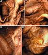
Endoscopic investigation of the internal organs of a 15th-century child mummy from Yangju, Korea
- PMID: 17062024
- PMCID: PMC2100347
- DOI: 10.1111/j.1469-7580.2006.00637.x
Endoscopic investigation of the internal organs of a 15th-century child mummy from Yangju, Korea
Abstract
Our previous reports on medieval mummies in Korea have provided information on their preservation status. Because invasive techniques cannot easily be applied when investigating such mummies, the need for non-invasive techniques incurring minimal damage has increased among researchers. Therefore, we wished to confirm whether endoscopy, which has been used in non-invasive and minimally invasive studies of mummies around the world, is an effective tool for study of Korean mummies as well. In conducting an endoscopic investigation on a 15th-century child mummy, we found that well-preserved internal organs remained within the thoracic, abdominal and cranial cavities. The internal organs - including the brain, spinal cord, lung, muscles, liver, heart, intestine, diaphragm and mesentery - were easily investigated by endoscopy. Even the stool of the mummy, which accidentally leaked into the abdominal cavity during an endoscopic biopsy, was clearly observed. In addition, unusual nodules were found on the surface of the intestines and liver. Our current study therefore showed that endoscopic observation could provide an invaluable tool for the palaeo-pathological study of Korean mummies. This technique will continue to be used in the study of medieval mummy cases in the future.
Figures


Similar articles
-
Three-dimensional reconstruction of medieval child mummy in Yangju, Korea, using multi-detector computed tomography.Ann Anat. 2007;189(6):558-68. doi: 10.1016/j.aanat.2007.03.005. Ann Anat. 2007. PMID: 18077999
-
Paleoparasitological report on the stool from a Medieval child mummy in Yangju, Korea.J Parasitol. 2007 Jun;93(3):589-92. doi: 10.1645/GE-905R3.1. J Parasitol. 2007. PMID: 17626351
-
Ultramicroscopic study on the hair of newly found 15th century mummy in Daejeon, Korea.Ann Anat. 2006 Sep;188(5):439-45. doi: 10.1016/j.aanat.2006.03.006. Ann Anat. 2006. PMID: 16999207
-
Mummies.Am J Phys Anthropol. 2007;Suppl 45:162-90. doi: 10.1002/ajpa.20728. Am J Phys Anthropol. 2007. PMID: 18046750 Review.
-
[Buddhist mummies in Japan].Kaibogaku Zasshi. 1993 Aug;68(4):381-98. Kaibogaku Zasshi. 1993. PMID: 8237299 Review. Japanese.
Cited by
-
Mummification in Korea and China: Mawangdui, Song, Ming and Joseon Dynasty Mummies.Biomed Res Int. 2018 Sep 13;2018:6215025. doi: 10.1155/2018/6215025. eCollection 2018. Biomed Res Int. 2018. PMID: 30302339 Free PMC article. Review.
-
The potential for non-invasive study of mummies: validation of the use of computerized tomography by post factum dissection and histological examination of a 17th century female Korean mummy.J Anat. 2008 Oct;213(4):482-95. doi: 10.1111/j.1469-7580.2008.00955.x. J Anat. 2008. PMID: 19014355 Free PMC article.
-
Anatomical confirmation of computed tomography-based diagnosis of the atherosclerosis discovered in 17th century Korean mummy.PLoS One. 2015 Mar 27;10(3):e0119474. doi: 10.1371/journal.pone.0119474. eCollection 2015. PLoS One. 2015. PMID: 25816014 Free PMC article.
-
Radiological diagnosis of congenital diaphragmatic hernia in 17th century Korean mummy.PLoS One. 2014 Jul 2;9(7):e99779. doi: 10.1371/journal.pone.0099779. eCollection 2014. PLoS One. 2014. PMID: 24988465 Free PMC article.
-
Magnetic resonance imaging performed on a hydrated mummy of medieval Korea.J Anat. 2010 Mar;216(3):329-34. doi: 10.1111/j.1469-7580.2009.01185.x. Epub 2010 Jan 7. J Anat. 2010. PMID: 20070429 Free PMC article.
References
-
- Cesarani F, Martina MC, Ferraris A, et al. Whole-body three-dimensional multidetector CT of 13 Egyptian human mummies. Am J Roentgenol. 2003;180:597–606. - PubMed
-
- Cesarani F, Martina MC, Grilletto R, et al. Facial reconstruction of a wrapped Egyptian mummy using MDCT. Am J Roentgenol. 2004;183:755–758. - PubMed
-
- Gaafar H, Abdel-Monem MH, Elsheikh S. Nasal endoscopy and CT study of Pharaonic and Roman mummies. Acta Otolaryngol. 1999;119:257–260. - PubMed
-
- Hagedorn HG, Zink A, Szeimies U, Nerlich AG. Macroscopic and endoscopic examinations of the head and neck region in ancient Egyptian mummies. HNO. 2004;52:413–422. - PubMed
Publication types
MeSH terms
LinkOut - more resources
Full Text Sources

