. 2006;34(20):5829-38.
doi: 10.1093/nar/gkl708. Epub 2006 Oct 24.

Structural basis of the Methanothermobacter thermautotrophicus MCM helicase activity

Affiliations

Structural basis of the Methanothermobacter thermautotrophicus MCM helicase activity

Alessandro Costa et al. Nucleic Acids Res. 2006.

Abstract

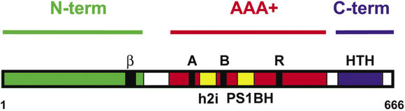
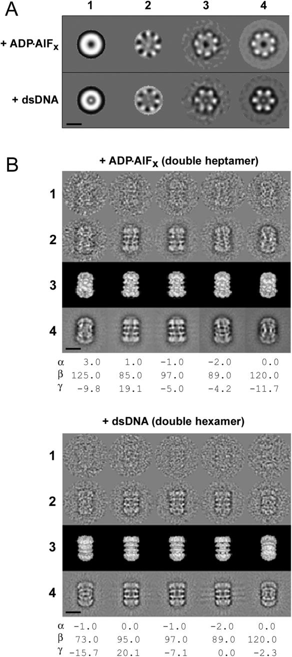
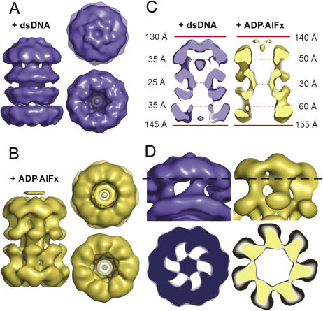
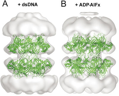
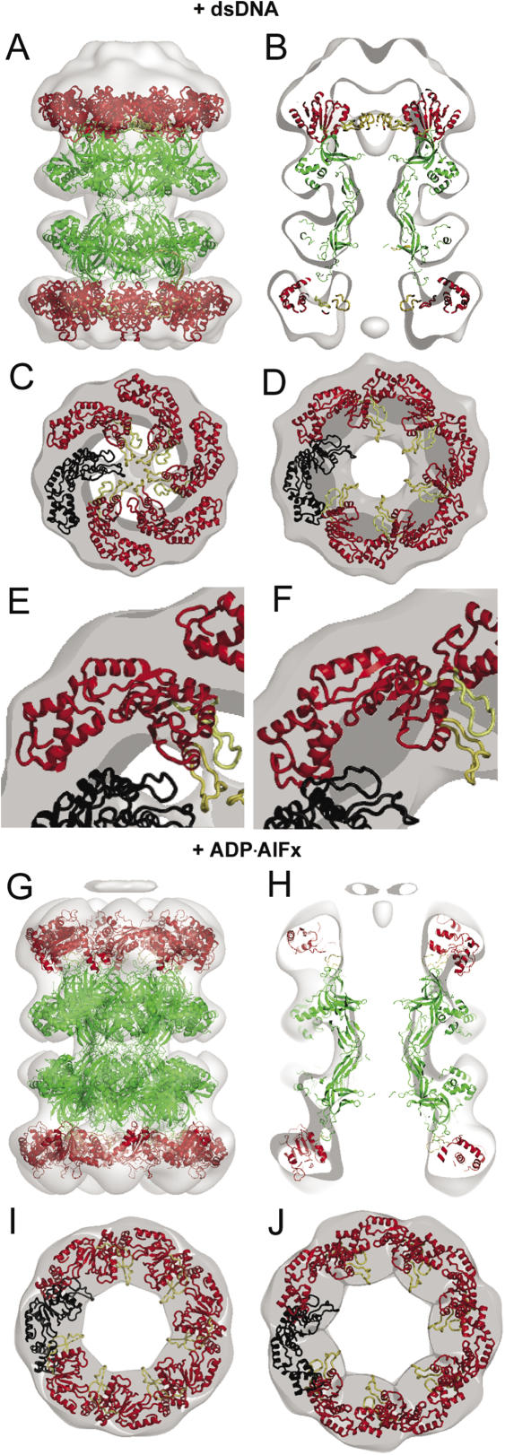
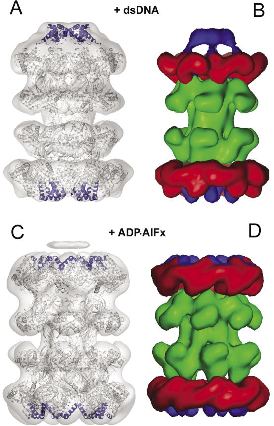
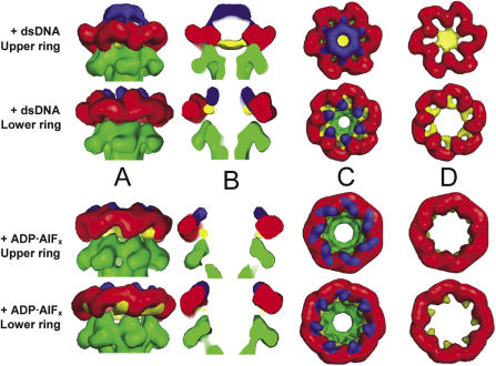
The MCM complex from the archaeon Methanother-mobacter thermautotrophicus is a model for the eukaryotic MCM2-7 helicase. We present electron-microscopy single-particle reconstructions of a DNA treated M.thermautotrophicus MCM sample and a ADP.AlF(x) treated sample, respectively assembling as double hexamers and double heptamers. The electron-density maps display an unexpected asymmetry between the two rings, suggesting that large conformational changes can occur within the complex. The structure of the MCM N-terminal domain, as well as the AAA+ and the C-terminal HTH dom-ains of ZraR can be fitted into the reconstructions. Distinct configurations can be modelled for the AAA+ and the HTH domains, suggesting the nature of the conformational change within the complex. The pre-sensor 1 and the helix 2 insertions, important for the activity, can be located pointing towards the centre of the channel in the presence of DNA. We propose a mechanistic model for the helicase activity, based on a ligand-controlled rotation of the AAA+ subunits.


Figures

Figure 1
Figure 1
Domain structure of MthMCM. The 242 amino acid N-terminal domain (green) contains a β-hairpin (β) capable of interacting with DNA. The central AAA+ domain (red) contains a Walker A, Walker B and arginine finger (R) motifs typical of AAA+ ATPases. In addition it contains a pre-sensor 1 β-hairpin (PS1BH) insertion and a helix 2 insertion (h2i), highlighted in yellow. The C-terminal domain contains a predicted helix–turn–helix motif (HTH, blue).
Figure 2
Figure 2
Single-particle analysis and 3D reconstruction. (A) Single-particle analysis performed on end-on views (21): (1) rotationally averaged total sum of end-on views; (2) most representative eigen images from end-on views, showing the symmetry component obtained performing MSA symmetry analysis; (3) characteristic end-on class averages obtained summing 100 images; (4) symmetry-imposed class averages. In the dsDNA treated protein, the particles present an intense peak of electron-density in the centre of the ring, which is lacking in the absence of DNA. This feature is particularly striking both in the rotationally averaged total sum and in the class averages. (B) Overview of the 3D reconstruction procedure of the double-hexameric and the double-heptameric structures (1) Examples of original images of MthMCM stained with uranyl acetate. These images are members of the class averages shown in the row below (protein is white). (2) Class averages (characteristic views) obtained by multi-reference alignment and classification. (3) Surface representations of the 3D reconstruction viewed from directions identical to the Euler directions assigned to the corresponding class averages in 2. (4) Reprojections of the 3D structure in the Euler-angle directions found for the class averages in 2. Scale bar 100 Å. While carrying out the reconstruction, the pertinent symmetry was imposed according to the indication obtained from MSA symmetry analysis carried out on ring-shaped particles (end-on views, A). According to previous single-particle analysis studies (21), >90% of the particle population is consistent with the formation of double heptamers in the presence of ADP·AlFx and >80% of the particle population forms a double hexamer when treated with DNA.
Figure 3
Figure 3
3D reconstructions of MthMCM complexes. Surface rendering of (A) the DNA treated MCM double hexamer (blue) and (B) the ADP·AlFx treated double heptamer (yellow), showing a head-to-head double-ring configuration with lateral holes. The two rings display an asymmetry, which is more pronounced in the double hexamer. (C) Slice-through side views of the double hexamer (blue) and the double heptamer (yellow), showing the internal channel. The external diameter of the upper and lower rings (continuous red line) and the diameter of the channel (dotted red line) are shown on the sides for each of the reconstructions. The upper ring in the double hexamer shows the presence of electron density in the centre of the channel. The double heptamer has no large peak of electron density inside the channel, and wide chambers (50–60 Å) in the centre of the two rings. (D) A tilted view of the upper hexameric ring (blue) shows a single lateral hole per subunit; a section of the ring (at the level indicated by the dashed line) illustrates the presence of elongated features departing from the protein walls and pointing inside the central channel. A side-view of the upper heptameric ring (yellow) shows a lateral hole divided in two apertures by the presence of an isthmus of electron density, while a section emphasises the lack of any electron density pointing inside the central channel.
Figure 4
Figure 4
Fitting of the MthMCM N-terminal domain. (A) Side-view of the atomic coordinates of the double hexameric N-terminal domain (5) (green) fitted into the electron-density map of the DNA treated sample. (B) A modelled heptameric N-terminal domain fitted into the electron density of the ADP·AlFx treated sample. The good fit to the electron density suggests that the head-to-head symmetry shown by the crystal structure is also maintained in the single-particle reconstruction of the full-length protein.
Figure 5
Figure 5
Fitting of the AAA+ ATPase domain of the ZraR transcriptional activator. The atomic coordinates of ZraR model derived from the crystal structure (32) were fitted to the AAA+ modules of MCM. The AAA+ domain of ZraR is shown in red, whereas the N-terminal domain of MthMCM is shown in green. One AAA+ subunit is colored in black. (A) Side-view of the double hexamer. (B) Side-view section of the double hexamer. (C) Section through the top ring of the double hexamer. (D) Section through the bottom ring of the double hexamer. (E) Close-up of the section of the top ring shown in (C). The h2i and PS1BH (shown in yellow) are pointing into the central channel and account for the electron density. (F) Close-up of the section of the bottom ring shown in D. The h2i and PS1BH (shown in yellow) pack against the ring subunits. (G) Side-view of the double heptamer. (H) Side-view section of the double hexamer. (I) Section through the top ring of the double heptamer. (J) Section through the bottom ring of the double heptamer. In the model of the double heptamer, the h2i and PS1BH pack between subunits in both the upper and lower rings.
Figure 6
Figure 6
Proposed location of the C-terminal domain. (A) The crystal structure of the HTH motif from the C-terminus of ZraR (blue) fitted into the 3D reconstruction of the double hexamer. Different relative positions of the C-terminal domain and AAA+ domains have been used to fit the upper and lower rings. (B) Surface representation obtained by filtering to 20 Å resolution the electron density calculated from the modelled atomic structures. The N-terminal domain is shown in green, the AAA+ domain in red and C-terminal domain in blue. (C) The crystal structure of the HTH motif from the C-terminus of ZraR (blue) fitted into the 3D reconstruction of the double heptamer. The AAA+ and C-terminal domains are in the same relative orientation as in the lower hexameric ring. (D) Surface representation of the heptameric model calculated and coloured as in (B).
Figure 7
Figure 7
Surface representation of the conformational changes occurring within the MthMCM rings. The N-terminal domain is shown in green, the AAA+ domain in red, the PS1BH and h2i insertions in yellow and the C-terminal domain in blue. To simplify the comparison with the upper rings, the lower rings have been inverted. The structures are viewed from (A) the side, (B) a slice through the side, (C) the top and (D) the top after the N- and C-terminal domains have been removed. The conformational changes within the upper and lower rings of the double hexamer are reminiscent of the nucleotide-controlled iris-type motion described for the LtAg helicase (25). Such a movement involves both the AAA+ and the C-terminal domain. Due to a rotation of the AAA+ domain, the PS1BH and h2i insertions move from pointing inside the channel to an inter-subunit position. Concertedly, the C-terminal domain rearranges from forming a distinct cap on top of the AAA+ tier to a less extended configuration.

References

    1. Chong J.P. Learning to unwind. Nature Struct. Mol. Biol. 2005;12:734–736. - PubMed
    1. Chong J.P., Hayashi M.K., Simon M.N., Xu R.M., Stillman B. A double-hexamer archaeal minichromosome maintenance protein is an ATP-dependent DNA helicase. Proc. Natl Acad. Sci. USA. 2000;97:1530–1535. - PMC - PubMed
    1. Shechter D.F., Ying C.Y., Gautier J. The intrinsic DNA helicase activity of Methanobacterium thermoautotrophicum delta H minichromosome maintenance protein. J. Biol. Chem. 2000;275:15049–15059. - PubMed
    1. Kelman Z., Lee J.K., Hurwitz J. The single minichromosome maintenance protein of Methanobacterium thermoautotrophicum DeltaH contains DNA helicase activity. Proc. Natl Acad. Sci. USA. 1999;96:14783–14788. - PMC - PubMed
    1. Fletcher R.J., Bishop B.E., Leon R.P., Sclafani R.A., Ogata C.M., Chen X.S. The structure and function of MCM from archaeal M. thermoautotrophicum. Nature Struct. Biol. 2003;10:160–167. - PubMed

Publication types

MeSH terms