. 2007 Feb 15;109(4):1533-40.
doi: 10.1182/blood-2006-08-040196. Epub 2006 Oct 24.

The Slc35d3 gene, encoding an orphan nucleotide sugar transporter, regulates platelet-dense granules

Affiliations

The Slc35d3 gene, encoding an orphan nucleotide sugar transporter, regulates platelet-dense granules

Sreenivasulu Chintala et al. Blood. .

Abstract

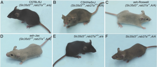
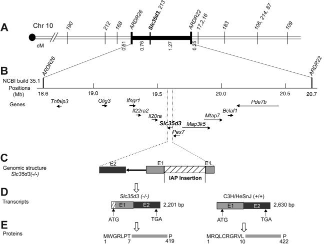
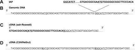
Platelet dense granules are lysosome-related organelles which contain high concentrations of several biologically important low-molecular-weight molecules. These include calcium, serotonin, adenine nucleotides, pyrophosphate, and polyphosphate, which are necessary for normal blood hemostasis. The synthesis of dense granules and other lysosome-related organelles is defective in inherited diseases such as Hermansky-Pudlak syndrome (HPS) and Chediak-Higashi syndrome (CHS). HPS and CHS mutations in 8 human and at least 16 murine genes have been identified. Previous studies produced contradictory findings for the function of the murine ashen (Rab27a) gene in platelet-dense granules. We have used a positional cloning approach with one line of ashen mutants to establish that a new mutation in a second gene, Slc35d3, on mouse chromosome 10 is the basis of this discrepancy. The platelet-dense granule defect is rescued in BAC transgenic mice containing the normal Slc35d3 gene. Thus, Slc35d3, an orphan member of a nucleotide sugar transporter family, specifically regulates the contents of platelet-dense granules. Unlike HPS or CHS genes, it has no apparent effect on other lysosome-related organelles such as melanosomes or lysosomes. The ash-Roswell mouse mutant is an appropriate model for human congenital-isolated delta-storage pool deficiency.


Figures

Figure 1
Figure 1
Effects of the Slc35d3, Rab27a, and agouti (A) genes on pigmentation. Control C57BL/6J (A) and C3H/HeSnJ (B) mice display typical nonagouti (a) and agouti (A) pigmentation, respectively. The ash-Roswell (C) and ash-Jax (D) mutants, which arose on the C3H/HeSnJ inbred line, are hypopigmented because of the homozygous Rab27a (ashen) mutation. The Slc35d3 mutation does not affect coat or eye color when transferred in homozygous form to the C57BL/6J (E) or C3H/HeSnJ (F) backgrounds.
Figure 2
Figure 2
Expression of Rab27a and Rab27b proteins in platelets of ash-Roswell and ash-Jax mutants. Platelet protein (30 μg) was electrophoresed and immunoblotted with the indicated antibodies. β-Actin served as loading control. The vertical dotted lines indicate that empty spacer gel lanes were removed during the digital composition of this figure.
Figure 3
Figure 3
Identification of the Slc35d3 gene by positional cloning. (A) High-resolution genetic map of the region surrounding the Slc35d3 gene on mouse chromosome 10. Numbered markers are polymorphic MIT microsatellites. ARDR26 (amplified with primers 5′-GGGTGGAGCGGTACTATTCA-3′ and 5′-TGTCTTCAGAGACAACAATCCAG-3′) and ARDR22 (amplified with primers 5′-TTTTCTTTAGGTGGTTTTCTACAGC-3′ and 5′-AGGACAGACAGAGCCCTGAG-3′) are polymorphic microsatellites identified by genome scanning. (B) High-resolution physical map [based on the National Center for Biotechnology Information map viewer (build 35.1)]. The positions of 11 known genes in the critical genetic interval are indicated with their transcription orientations indicated by arrows. (C) The region of the genome from the beginning of exon 1 to the end of exon 2 of Slc35d3. A 5.5-kb IAP element is inserted between nucleotides 12296009 and 12296010 (Contig NT_039 492.5 of Mus musculus build 35.1) in exon 1 of Slc35d3 in the ash-Roswell mutant (Slc35d3−/−). (D) Transcripts of Slc35d3 in ash-Roswell (Slc35d3−/−) and control C3H/HeSnJ brains. Slc35d3 m-RNA of ash-Roswell mutants contains a new and in-frame ATG start site derived from the IAP element. More detailed views of normal and mutant Slc35d3 transcripts are given in Figure 4. (E) Predicted N-terminal sequences of mutant and control Slc35d3 proteins. The substitution of 7 new N-terminal mutant amino acids for the original 10 N-terminal wild-type amino acids is indicated.
Figure 4
Figure 4
Genomic and transcript structures of Slc35d3 in ash-Roswell and C3H/HeSnJ. Insertion of an IAP element (A) into exon 1 of the Slc35d3 gene (B) alters the 5′ terminal sequence of the Slc35d3 cDNA (C) to introduce a new IAP-derived ATG start site (bold and underlined in panel C) in the ash-Roswell mutant. This results in substitution of 21 new in-frame coding nucleotides (parentheses in panel C) for the 30 coding nucleotides (parentheses in D) found in control C3H DNA. The predicted result is the substitution of 7 new N-terminal amino acids in mutant Slc35d3 (see Figure 3E). IAP-derived sequences are in bold. A 7-bp duplication of endogenous gene sequences (GGCATCT), which is typical of IAP transpositions, is underlined. ATG start signals derived from the IAP and from the Slc35d3 gene are underlined. An additional 420 nucleotides at the 5′ end of the C3H/HeSnJ wild-type Slc35d3 cDNA are not listed.
Figure 5
Figure 5
Relative expression of Slc35d3 in control and mutant tissues. (A-B) Multiple tissue Northern blots (Clontech) were treated with a 303-bp cDNA probe derived from nucleotides 1281 to 1583 of Slc35d3 cDNA together with a β-actin probe (below) as a loading control. Poly(A)–RNA was isolated from Melan-A melanocytes and C3H/HeSnJ brain for Northern blotting (C). Brain and spleen poly(A)-RNA blots (4 μg/lane) (D) were probed with the above Slc35d3 probe and a G3PDH probe (below). The apparent decreased expression of the 2.6-kb mRNA in control C3H brain tissue in this experiment compared with that of control brain tissue in panel C is due to the greatly decreased time (8 hours compared with 4 days) of exposure of this blot to film.
Figure 6
Figure 6
Platelet serotonin concentrations in BAC-positive and BAC-negative F2 progeny. Platelet serotonin levels were determined in each of 22 BAC-positive, Slc35d3(−/− or +/−) (●), 9 BAC-negative, Slc35d3−/− (■), and 5 BAC-negative Slc35d3+/− (▴) F2 progeny. Each symbol represents the serotonin concentration in platelets from a single mouse. The average serotonin levels in wild-type C3H/HeSnJ (solid arrow) and ash-Roswell mutants (open arrow) are indicated.

References

    1. Rendu F, Brohard-Bohn B. The platelet release reaction: granules' constituents, secretion and functions. Platelets. 2001;12:261–273. - PubMed
    1. McNicol A, Israels SJ. Platelet dense granules: structure, function and implications for haemostasis. Thromb Res. 1999;95:1–18. - PubMed
    1. Ruiz FA, Lea CR, Oldfield E, Docampo R. Human platelet-dense granules contain polyphosphate and are similar to acidocalcisomes of bacteria and unicellular eukaryotes. J Biol Chem. 2004;279:44250–44257. - PubMed
    1. Smith SA, Mutch NJ, Baskar D, Rohloff P, Docampo R, Morrissey JH. Polyphosphate modulates blood coagulation and fibrinolysis. Proc Natl Acad Sci U S A. 2006;103:903–908. - PMC - PubMed
    1. Youssefian T, Cramer EM. Megakaryocyte dense granule components are sorted in multivesicular bodies. Blood. 2000;95:4004–4007. - PubMed

Publication types

Substances