. 2007 Feb 15;109(4):1433-41.
doi: 10.1182/blood-2006-06-031898. Epub 2006 Oct 24.

The megakaryocyte lineage originates from hemangioblast precursors and is an integral component both of primitive and of definitive hematopoiesis

Affiliations

The megakaryocyte lineage originates from hemangioblast precursors and is an integral component both of primitive and of definitive hematopoiesis

Joanna Tober et al. Blood. .

Abstract

In the adult, platelets are derived from unipotential megakaryocyte colony-forming cells (Meg-CFCs) that arise from bipotential megakaryocyte/erythroid progenitors (MEPs). To better define the developmental origin of the megakaryocyte lineage, several aspects of megakaryopoiesis, including progenitors, maturing megakaryocytes, and circulating platelets, were examined in the murine embryo. We found that a majority of hemangioblast precursors during early gastrulation contains megakaryocyte potential. Combining progenitor assays with immunohistochemical analysis, we identified 2 waves of MEPs in the yolk sac associated with the primitive and definitive erythroid lineages. Primitive MEPs emerge at E7.25 along with megakaryocyte and primitive erythroid progenitors, indicating that primitive hematopoiesis is bilineage in nature. Subsequently, definitive MEPs expand in the yolk sac with Meg-CFCs and definitive erythroid progenitors. The first GP1bbeta-positive cells in the conceptus were identified in the yolk sac at E9.5, while large, highly reticulated platelets were detected in the embryonic bloodstream beginning at E10.5. At this time, the number of megakaryocyte progenitors begins to decline in the yolk sac and expand in the fetal liver. We conclude that the megakaryocyte lineage initially originates from hemangioblast precursors during early gastrulation and is closely associated both with primitive and with definitive erythroid lineages in the yolk sac prior to the transition of hematopoiesis to intraembryonic sites.


Figures
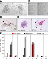
Figure 1
Figure 1
Megakaryocyte potential is derived from hemangioblast precursors. (A) Hemangioblast-derived colony from E7.5 mouse embryo (i). Adherent and nonadherent cells from a hemangioblast-derived colony after 3 days in liquid expansion media (ii). (B) Proplatelet formation of cells differentiated from a hemangioblast-derived colony after 3 days in liquid media (i-ii). (C) PECAM-positive cells (arrowheads) in the adherent cell population after 10 days in liquid expansion media. (D) Primitive erythroid and megakaryocyte colony formation from expanded, nonadherent cells placed into progenitor assays. An EryP-CFC–derived colony contains cells positive for βH1-globin (i) and a Meg-CFC–derived colony contains cells and proplatelet extensions (arrowheads) positive for GP1bβ (ii). (E) Hematopoietic potential of 10 individual hemangioblast colonies expanded in liquid culture. The number of EryP-CFCs, Meg-CFCs, and megakaryocyte cells (meg cells) is shown. Scale bars represent 10 μm.
Figure 2
Figure 2
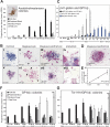
Meg-CFCs and 2 novel bipotential MEPs emerge during embryonic hematopoiesis. (A) Number (+SEM) of acetylcholinesterase-positive (megakaryocyte) colonies grown from individual embryonic tissues by stage. The inset (i) shows an acetylcholinesterase-positive colony. (B) Immunohistochemical staining of erythroid and megakaryocyte progenitor–derived colonies. (Top) Primitive erythroid (i), megakaryocyte (ii), and bipotential primitive MEP (iii) colonies from neural plate–stage cultures stained with primitive erythroid-specific βH1-globin (blue) and megakaryocyte-specific GP1bβ (pink) antibodies. (Bottom) Erythroid (v), megakaryocyte (vi), and bipotential definitive MEP (vii) colonies from E9.5 to E10.5 yolk sac stained with panerythroid Ter119 (blue) and with GP1bβ (pink) antibodies. Boxed areas in panels iii and vii highlight proplatelet formation at 100 × shown in panels iv and viii, respectively. Arrowheads indicate proplatelet formation in panels ii and vi. Scale bars represent 10 μm. (C) Average number (+SEM) of primitive erythroid, megakaryocyte, and bipotential primitive MEP colonies from plated whole embryos (PS-LNP) and yolk sac (HF-46 sp) as determined by immunohistochemistry. (D) Primitive MEP–derived colony derived from a neural plate–staged embryo contains cells positive for βH1-globin and CD41. (E) Number (+SEM) of βH1-globin/GP1bβ-double positive colonies from a dilution series of 4 to 5 sp yolk sacs, in triplicate. (F) Spatial and temporal distribution of Meg-CFCs as determined by GP1bβ-stained colonies per tissue. (G) Spatial and temporal distribution of definitive MEPs, as determined by Ter119/GP1bβ double-positive colonies per tissue. sp indicates somite pair; PS, preprimitive streak; MS, mid primitive streak; LS, late primitive streak; ENP, early neural plate; MNP, mid neural plate; LNP, late neural plate; HF, head fold; and LHF, late head fold. Approximate embryonic day (E) is provided below the developmental stages.
Figure 3
Figure 3
Enrichment of progenitors in extraembryonic (yolk sac) versus intraembryonic regions. Numbers of primitive MEP, definitive MEP, or unipotential Meg-CFC hematopoietic progenitors (as determined by immunohistochemistry) and erythroblasts in all intraembryonic tissues versus yolk sac. Fold enrichment was calculated as the ratio of intraembryonic progenitors to erythroblasts divided by the ratio of yolk sac progenitors to erythroblasts. Tissues from individual experiments were grouped and averaged by stage (± SEM)..
Figure 4
Figure 4
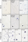
Localization of megakaryocytes (GP1bβ-positive cells) in mouse embryos. Individual megakaryocytes were first identified in the yolk sac of E9.5 conceptuses (arrows, top row, left panel). No GP1bβ-positive cells were detected in the E9.5 embryo proper (top row, middle panel). Maternally derived GP1bβ-positive platelets were detected in the surrounding E9.5 decidua (top, right panel). At E10.5 (middle row), clusters of GP1bβ-positive cells were evident in the yolk sac and intraembryonic megakaryocytes were seen in the circulation (arrowhead), associated with the wall of the aorta (arrows), and in the fetal liver (arrowhead). GP1bβ antibodies label both small (open arrowhead) and large cells in the E11.5 to E12.5 fetal liver, and predominantly large cells in the E13.5 to E14.5 fetal liver (lower row). Platelets are seen in E11.5 to 14.5 livers (arrows). Scale bars represent 50 μm.
Figure 5
Figure 5
Large platelets circulate in the embryonic and fetal vasculature. (A) Platelet-enriched blood was stained with anti–GPV-FITC antibodies (E11.5 shown, left panel), which specifically label platelets but not red blood cells. Shown to the right are examples of platelets from E11.5, E15.5, and adult peripheral blood, depicting the broad face (top) and narrow side (bottom). Regardless of cell diameter, all platelets were narrow and biconvex. Scale bar equals 5 μm. (B) Mean platelet diameter (± SEM) in E11.5 to E15.5 fetuses, in neonates, and in adult mice. A minimum of 100 platelets was measured per peripheral blood sample and at least 3 independent samples were studied, except for E10.5, of which 2 pooled blood samples were examined and a total of 11 platelets was measured. (C) Transmission electron microscopic images of E11.5, E15.5, and adult platelets. All images taken at × 10 000. Open canalicular system (arrows), dense granules (arrowheads), and alpha granules (α) are evident.
Figure 6
Figure 6
Fetal platelets are highly reticulated. (A) Flow cytometric analysis of adult and fetal blood. Platelet fractions were identified by forward and side scatter parameters (left panels), lack of Ter119 and Hoechst staining (middle panels), and CD41 staining (right panels). Thiazole orange–positive platelets were gated as described in “Materials and methods.” (B) The mean percentage (+SEM) of reticulated platelets in E11.5 to E15.5 fetuses (■), in maternal blood (▩), in neonates (▤), and in nonpregnant adult mice (□). Fetal blood samples were analyzed from 3 littermates (E11.5, E14.5, and neonate), 3 fetuses from 3 separate experiments (E12.5), 4 embryos from 2 separate experiments (E13.5), or taken from a single fetus (E15.5). Adult blood was analyzed from a single sample at gestational days 13.5 to 15.5, in triplicate at gestational days 11.5 to 12.5, and from 5 independent nonpregnant adult samples. (C) Mean fluorescence (± SEM) of thiazole orange in untreated platelets and platelets treated with 10 μg/mL RNase A.

References

    1. Moore MA, Metcalf D. Ontogeny of the haemopoietic system: yolk sac origin of in vivo and in vitro colony forming cells in the developing mouse embryo. Br J Haematol. 1970;18:279–296. - PubMed
    1. Sabin FR. Studies on the origin of blood vessels and red blood corpuscles as seen in the living blastoderm of chicks during the second day of incubation. Contrib Embryol. 1920;9:213–262.
    1. Choi K, Kennedy M, Kazarov A, Papadimitriou JC, Keller G. A common precursor for hematopoietic and endothelial cells. Development. 1998;125:725–732. - PubMed
    1. Nishikawa SI, Nishikawa S, Hirashima M, Matsuyoshi N, Kodama H. Progressive lineage analysis by cell sorting and culture identifies FLK1+VE-cadherin+ cells at a diverging point of endothelial and hemopoietic lineages. Development. 1998;125:1747–1757. - PubMed
    1. Lacaud G, Gore L, Kennedy M, et al. Runx1 is essential for hematopoietic commitment at the hemangioblast stage of development in vitro. Blood. 2002;100:458–466. - PubMed

Publication types