

Craniofacial tissue engineering by stem cells
- PMID: 17062735
- PMCID: PMC2571078
- DOI: 10.1177/154405910608501101
Craniofacial tissue engineering by stem cells
Abstract
Craniofacial tissue engineering promises the regeneration or de novo formation of dental, oral, and craniofacial structures lost to congenital anomalies, trauma, and diseases. Virtually all craniofacial structures are derivatives of mesenchymal cells. Mesenchymal stem cells are the offspring of mesenchymal cells following asymmetrical division, and reside in various craniofacial structures in the adult. Cells with characteristics of adult stem cells have been isolated from the dental pulp, the deciduous tooth, and the periodontium. Several craniofacial structures--such as the mandibular condyle, calvarial bone, cranial suture, and subcutaneous adipose tissue--have been engineered from mesenchymal stem cells, growth factor, and/or gene therapy approaches. As a departure from the reliance of current clinical practice on durable materials such as amalgam, composites, and metallic alloys, biological therapies utilize mesenchymal stem cells, delivered or internally recruited, to generate craniofacial structures in temporary scaffolding biomaterials. Craniofacial tissue engineering is likely to be realized in the foreseeable future, and represents an opportunity that dentistry cannot afford to miss.
Figures

References
-
- About I, Bottero MJ, de Denato P, Camps J, Franquin JC, Mitsiadis TA. Human dentin production in vitro. Exp Cell Res. 2000;258:33–41. - PubMed
-
- Akizuki T, Oda S, Komaki M, Tsuchioka H, Kawakatsu N, Kikuchi A, et al. Application of periodontal ligament cell sheet for periodontal regeneration: a pilot study in beagle dogs. J Periodontal Res. 2005;40:245–251. - PubMed
-
- Alden TD, Beres EJ, Laurent JS, Engh JA, Das S, London SD, et al. The use of bone morphogenetic protein gene therapy in craniofacial bone repair. J Craniofac Surg. 2000;11:24–30. - PubMed
-
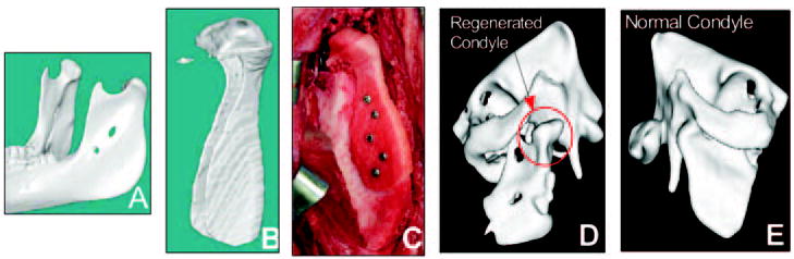
- Alhadlaq A, Mao JJ. Tissue-engineered neogenesis of human-shaped mandibular condyle from rat mesenchymal stem cells. J Dent Res. 2003;82:951–956. - PubMed
-
- Alhadlaq A, Mao JJ. Mesenchymal stem cells: isolation and therapeutics. Stem Cells Dev. 2004;13:436–448. - PubMed
Publication types
MeSH terms
Grants and funding
- R01 DE013028/DE/NIDCR NIH HHS/United States
- DE13964/DE/NIDCR NIH HHS/United States
- DE13194/DE/NIDCR NIH HHS/United States
- R01 DE012462/DE/NIDCR NIH HHS/United States
- R01 DE014526/DE/NIDCR NIH HHS/United States
- R29 DE012462/DE/NIDCR NIH HHS/United States
- R01 DE016530/DE/NIDCR NIH HHS/United States
- RC2 DE020767/DE/NIDCR NIH HHS/United States
- DE13397/DE/NIDCR NIH HHS/United States
- DE14526/DE/NIDCR NIH HHS/United States
- R01 DE013397/DE/NIDCR NIH HHS/United States
- DE013835/DE/NIDCR NIH HHS/United States
- EB02332/EB/NIBIB NIH HHS/United States
- DE16619/DE/NIDCR NIH HHS/United States
- R01 DE016129/DE/NIDCR NIH HHS/United States
- R01 DE013608/DE/NIDCR NIH HHS/United States
- R01 EB002332/EB/NIBIB NIH HHS/United States
- DE013608/DE/NIDCR NIH HHS/United States
- DE016129/DE/NIDCR NIH HHS/United States
- DE13028/DE/NIDCR NIH HHS/United States
- DE012462/DE/NIDCR NIH HHS/United States
- R21 DE016619/DE/NIDCR NIH HHS/United States
- R01 DE013964/DE/NIDCR NIH HHS/United States
- R01 DE015391/DE/NIDCR NIH HHS/United States
- DE15384/DE/NIDCR NIH HHS/United States
- DE007057/DE/NIDCR NIH HHS/United States
- R01 DE015384/DE/NIDCR NIH HHS/United States
- T32 DE007057/DE/NIDCR NIH HHS/United States
- R01 DE013835/DE/NIDCR NIH HHS/United States
- R01 DE013194/DE/NIDCR NIH HHS/United States
- DE016530/DE/NIDCR NIH HHS/United States
LinkOut - more resources
Full Text Sources
Other Literature Sources
Research Materials
