. 2006 Nov 21;103(47):17696-700.
doi: 10.1073/pnas.0607993103. Epub 2006 Oct 24.

A cryptochrome/photolyase class of enzymes with single-stranded DNA-specific photolyase activity

Affiliations

A cryptochrome/photolyase class of enzymes with single-stranded DNA-specific photolyase activity

Christopher P Selby et al. Proc Natl Acad Sci U S A. .

Abstract

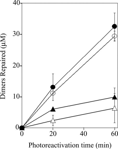
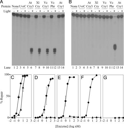
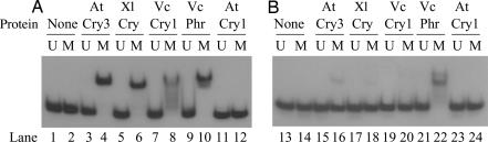
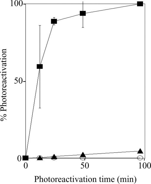
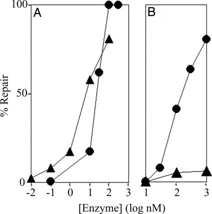
Photolyases and cryptochrome blue-light photoreceptors are evolutionarily related flavoproteins that perform distinct functions. Photolyases repair UV-damaged DNA in many species from bacteria to plants and animals. Cryptochromes regulate growth and development in plants and the circadian clock in animals. Recently, a new branch of the photolyase/cryptochrome family was identified. Members of this branch exhibited no or trace levels of DNA repair activity in vivo and in vitro and, therefore, were considered to be cryptochromes, and they were named cryptochrome-DASH. Here, we show that Cry-DASH proteins from bacterial, plant, and animal sources actually are photolyases with high degree of specificity for cyclobutane pyrimidine dimers in ssDNA.


Conflict of interest statement

The authors declare no conflict of interest.

Figures

Fig. 1.
Fig. 1.
Repair of cyclobutane pyrimidine dimers in poly dU(U<>U) and poly rU(U<>U). VcPhr or VcCry1 at a 100 nM concentration were mixed with a 100 μM concentration of rU<>rU or dU<>dU in the corresponding nucleotide polymers and exposed to photoreactivating light for the indicated time periods. The level of repair was determined from the increase in absorbance at 260 nm (16). ○, VcPhr with poly dU(U<>U) substrate; ●, VcCry1 with poly dU(U<>U) substrate. ▵, VcPhr with poly rU(U<>U) substrate; ▴, VcCry1 with poly rU(U<>U) substrate. Error bars denote SDs where they are larger than the symbols.
Fig. 2.
Fig. 2.
Repair of a cyclobutane thymine dimer (T<>T) in ss- and dsDNAs by photolyase and Cry-DASH family enzymes. (A and B) A 48-nt-long radiolabeled oligomer with a T<>T in the TTAA sequence (MseI recognition site) in the center, in single-stranded (A) or double-stranded (B) form, was mixed with the appropriate enzyme and exposed to photoreactivating light as indicated. The level of repair was determined by the susceptibility of the DNA to cleavage by MseI endonuclease. Single-stranded repair products were annealed with the complementary strand before digestion with MseI. The reaction products were separated on 5% polyacrylamide gels. Shown are autoradiograms of representative gels. Photolyase from Vibrio cholerae (VcPhr) was used as a positive control, and UvrC from Bacillus subtilis (UvrC) was used as a negative control. AtCry3, A. thaliana Cry-DASH; XlCry, Xenopus laevis Cry-DASH; VcCry1, V. cholerae Cry-DASH; AtCry1, A. thaliana cryptochrome blue-light photoreceptor. The substrate concentration was 0.27 nM, and the enzymes were at a concentration of 100 nM. Photoreactivation was for 2 h. (C–G) Repair of cyclobutane thymine dimer as a function of enzyme concentration for VcPhr (C), VcCry1 (D), XlCry (E), AtCry3 (F), and AtCry1 (G). Substrate at 0.27 nM was incubated with the indicated concentrations of enzymes and subjected to photoreactivating light for 2 h. ●, ssDNA; ▴, dsDNA. Error bars denote standard deviations where they are larger than the symbols.
Fig. 3.
Fig. 3.
Binding of VcPhr and Cry-DASH enzymes to a cyclobutane thymine dimer. A radiolabeled 48-mer, either unmodified (U) or modified (M) to contain a cyclobutane thymine dimer, was prepared in a single-stranded (A) or double-stranded (B) form. Substrates were incubated with the enzymes shown, and then the bound and free DNAs were separated with 5% polyacrylamide gels. Note that weak, apparent binding of AtCry3 and Xl-DASH to the dimer in dsDNA likely results from binding to contaminating ssDNA substrate as indicated by the loss of free ssDNA in lanes 16 and 18.
Fig. 4.
Fig. 4.
In vivo assay of photoreactivation by VcPhr and VcCry1 (Cry-DASH) enzymes. E. coli UNC523 (phruvrA) transformed with vectors expressing maltose binding protein (○), MBP-VcCry1 (▴), or MBP-VcPhr (■) was irradiated with 8 J·m−2 of 254 nm light to ≈2 × 10⁻⁵ survival. Cultures then were exposed to photoreactivating light (366 nm) at a fluence rate of 24 J·m−2·sec−1 for the indicated times and plated. Percent photoreactivation is plotted as a function of photoreactivation time. Error bars denote standard deviations where they are larger than the symbols.
Fig. 5.
Fig. 5.
Repair of UV-irradiated single- and double-stranded plasmids by VcPhr (A) and VcCry1 (Cry-DASH) (B) proteins. The plasmids were irradiated with 254 nm to generate approximately five photoproducts per plasmid as determined by the transformation assay (20). The irradiated plasmids were incubated with VcPhr or VcCry1 at the indicated concentrations, exposed to photoreactivating light for 2 h, and then used to transform E. coli strain CA1 (uvrA). From the number of transformants before and after light exposure, the percent dimers repaired by the photoenzyme was calculated (20). ●, single-stranded plasmid; ▴, double-stranded plasmid. Data points are from a representative experiment. Note that some subtle differences exist between the repair of single- and double-stranded plasmid DNA in relation to the T<>T in the single-stranded and double-stranded oligomers (Fig. 1). We presume that these differences reflect the heterogeneity of the photodimers in the plasmid substrates and the nature of the assays used to measure repair of the plasmids and the oligonucleotides.

References

    1. Cashmore AR. Cell. 2003;122:537–543. - PubMed
    1. Lin C, Shalitin D. Annu Rev Plant Biol. 2003;54:469–496. - PubMed
    1. Sancar A. Chem Rev. 2003;103:2203–2237. - PubMed
    1. Ahmad M, Cashmore AR. Nature. 1993;366:162–166. - PubMed
    1. Guo H, Yang H, Mockler TC, Lin C. Science. 1998;279:1360–1363. - PubMed

Publication types