

Space occupying lesions of parotid gland. Comparative diagnostic imaging and pathological analysis of echo color/power Doppler and of magnetic resonance imaging
- PMID: 17063984
- PMCID: PMC2639964
Space occupying lesions of parotid gland. Comparative diagnostic imaging and pathological analysis of echo color/power Doppler and of magnetic resonance imaging
Abstract
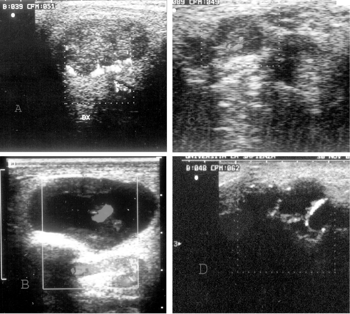
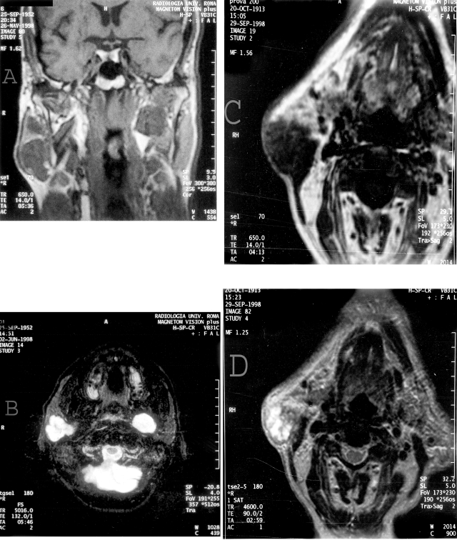
Aim of the study was to evaluate the role of echo color/power Doppler and magnetic resonance imaging in the diagnosis of space occupying parotid lesions, in the attempt to establish criteria for differential diagnosis between benign and malignant types. The study population comprised 49 patients (23 male, 26 female), age range 30-85 years, with a space occupying lesion of the parotid gland. Each lesion was carefully studied with ultrasound integrated with colour/power doppler and magnetic resonance imaging; patients were then submitted to echo-guided needle-biopsy and surgical excision. A preliminary evaluation by means of ultrasound revealed the site, size of lesion, echostructure and borders. Echo colour/power Doppler and magnetic resonance imaging can play a very important role both in diagnosis and pre-operative surgical planning of parotid lesions, although cost and availability, the former to be recommended vs. magnetic resonance imaging, which is poorly accessible, expensive, and not always accepted by the patients.
Lo scopo del presente studio è valutare il ruolo dell’eco-color power doppler e della risonanza magnetica nella diagnosi delle lesioni occupanti spazio della ghiandola parotide, nel tentativo di stabilire i criteri per una diagnosi differenziale tra formazioni benigne e maligne.
Lo studio ha preso in considerazione 49 pazienti (23 maschi e 26 femmine), di età compresa tra 30 e 85 anni, che presentavano lesioni tumorali della parotide. Tali formazioni sono state valutate con eco-color power doppler e risonanza magnetica, diretta e dopo somministrazione endovenosa di mezzo di contrasto paramagnetico quindi sottoposte a biopsia eco-guidata e successivamente ad escissione chirurgica. La valutazione preliminare condotta con ecografia ha considerato la posizione della lesione, le dimensioni, l’ecostruttura ed i margini. L’eco-color power doppler e la risonanza magnetica giocano entrambe un ruolo importante nella diagnsoi e nella pianificazione preoperatoria delle lesioni tumorali della parotide. La risonanza magnetica, a differenza dell’eco-color power doppler, presenta costi più elevati, un più difficile accesso e minore tollerabilità da parte di alcuni pazienti.
Figures
Similar articles
-
The role of Echo Colour/Power Doppler and magnetic resonance in expansive parotid lesions.J Exp Clin Cancer Res. 2004 Dec;23(4):585-92. J Exp Clin Cancer Res. 2004. PMID: 15743028
-
Salivary gland lipomas: ultrasonographic and magnetic resonance imaging.J Craniofac Surg. 2007 Nov;18(6):1464-6. doi: 10.1097/scs.0b013e31814e056c. J Craniofac Surg. 2007. PMID: 17993902
-
[Doppler-color ultrasonography in the diagnosis of parotid tumors].Acta Otorhinolaryngol Ital. 1997 Feb;17(1):52-7. Acta Otorhinolaryngol Ital. 1997. PMID: 9412155 Clinical Trial. Italian.
-
Parotid Gland Lesions: Multiparametric Ultrasound and MRI Features.Ultraschall Med. 2016 Oct;37(5):454-471. doi: 10.1055/s-0042-109171. Epub 2016 Jun 14. Ultraschall Med. 2016. PMID: 27300273 Review. English.
-
Pitfalls in the staging of cancer of the major salivary gland neoplasms.Neuroimaging Clin N Am. 2013 Feb;23(1):107-22. doi: 10.1016/j.nic.2012.08.009. Neuroimaging Clin N Am. 2013. PMID: 23199664 Review.
Cited by
-
A prospective study of three diagnostic sonographic methods in differentiation between benign and malignant salivary gland tumours.Dentomaxillofac Radiol. 2011 Dec;40(8):476-85. doi: 10.1259/dmfr/18407834. Dentomaxillofac Radiol. 2011. PMID: 22065796 Free PMC article.
-
Improving the diagnosis of common parotid tumors via the combination of CT image biomarkers and clinical parameters.BMC Med Imaging. 2020 Apr 15;20(1):38. doi: 10.1186/s12880-020-00442-x. BMC Med Imaging. 2020. PMID: 32293304 Free PMC article.
-
Palatine Tonsil Measurements and Echogenicity during Tonsillitis Using Ultrasonography: A Case-Control Study.Diagnostics (Basel). 2023 Feb 15;13(4):742. doi: 10.3390/diagnostics13040742. Diagnostics (Basel). 2023. PMID: 36832230 Free PMC article.
References
-
- Yousem DM, Kraut MA, Chalian AA. Major salivary gland imaging. Radiology 2000;216:19-29. - PubMed
-
- Joe VQ, Westesson PL. Tumours of the parotid gland: MR imaging characteristics of various histologic types. Am J Roentgenol 1994;163:433-8. - PubMed
-
- Wolf JS, Goldberg AN, Bigelow DC. Pleomorphic adenoma of the parotid. Am Fam Physician 1997;56:185-92. - PubMed
-
- Grazioli L, Olivetti L, Matricardi L, Zanetti U, Burlini D, Negrini S, et al. Comparison of ultrasonography, computerized tomography, and magnetic resonance in the study of parotid. Radiol Med 1993;86:268-80. - PubMed
-
- Gritzmann N. Sonography of the salivary glands. Am J Roentgenol 1989;153:161-6. - PubMed
Publication types
MeSH terms
LinkOut - more resources
Full Text Sources
Medical