. 2006 Dec;99(5):1338-50.
doi: 10.1111/j.1471-4159.2006.04161.x. Epub 2006 Oct 25.

Calcitonin gene-related peptide enhances release of native brain-derived neurotrophic factor from trigeminal ganglion neurons

Affiliations

Calcitonin gene-related peptide enhances release of native brain-derived neurotrophic factor from trigeminal ganglion neurons

Ilya Buldyrev et al. J Neurochem. 2006 Dec.

Abstract

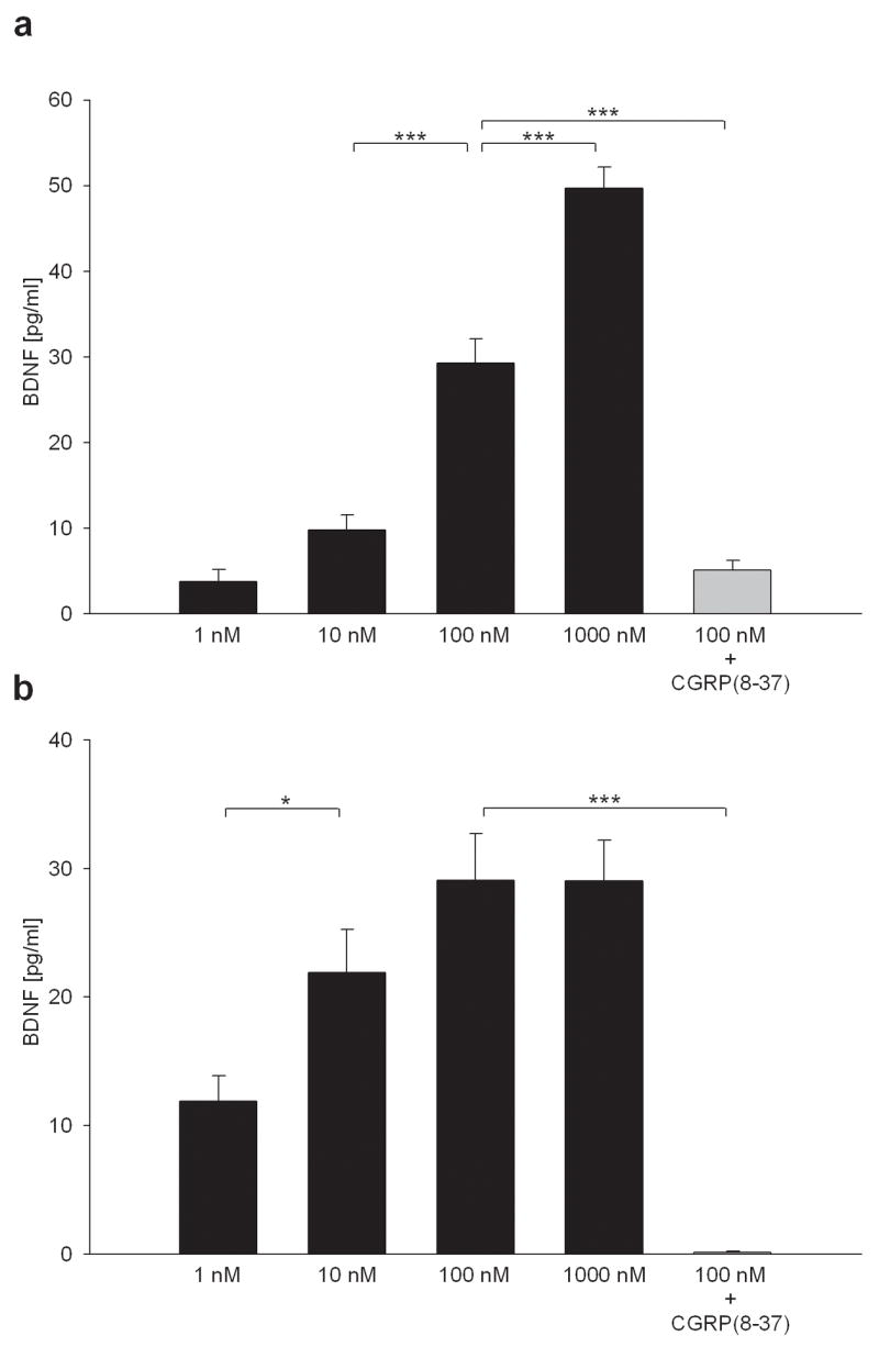
Activity-dependent plasticity in nociceptive pathways has been implicated in pathomechanisms of chronic pain syndromes. Calcitonin gene-related peptide (CGRP), which is expressed by trigeminal nociceptors, has recently been identified as a key player in the mechanism of migraine headaches. Here we show that CGRP is coexpressed with brain-derived neurotrophic factor (BDNF) in a large subset of adult rat trigeminal ganglion neurons in vivo. Using ELISA in situ, we show that CGRP (1-1000 nM) potently enhances BDNF release from cultured trigeminal neurons. The effect of CGRP is dose-dependent and abolished by pretreatment with CGRP receptor antagonist, CGRP(8-37). Intriguingly, CGRP-mediated BDNF release, unlike BDNF release evoked by physiological patterns of electrical stimulation, is independent of extracellular calcium. Depletion of intracellular calcium stores with thapsigargin blocks the CGRP-mediated BDNF release. Using transmission electron microscopy, our study also shows that BDNF-immunoreactivity is present in dense core vesicles of unmyelinated axons and axon terminals in the subnucleus caudalis of the spinal trigeminal nucleus, the primary central target of trigeminal nociceptors. Together, these results reveal a previously unknown role for CGRP in regulating BDNF availability, and point to BDNF as a candidate mediator of trigeminal nociceptive plasticity.


Figures

Figure 1
Figure 1
BDNF protein is expressed by a large subset of adult rat trigeminal ganglion (TG) neurons, including CGRP-immunoreactive cells. Micrographs of a single 8-μm section of an adult TG double-immunostained for BDNF (αBDNF) and CGRP (αCGRP). A large number of BDNF-immunoreactive cells can be seen throughout the section. An overlay image (αBDNF/αCGRP) shows that many CGRP-positive cells also express BDNF immunoreactivity (arrows). Scale bar 100 μm.
Figure 2
Figure 2
Endogenously expressed BDNF does not support survival of postnatal TG neurons in vitro. (a) A representative example of a neuron-enriched dissociate culture of newborn rat TG, double-immunostained for BDNF (αBDNF) and Neurofilament 68 and 160, a pan neuronal marker (αNF). Control cultures, in which the primary anti-BDNF antibody was omitted, were completely devoid of staining. Scale bar 50 μm. (b) Total number of NF-positive cells per culture in control conditions (Control), in the presence of TrkB-Fc fusion protein, an immunological agent that inhibits TrkB receptor activation by binding BDNF (TrkB-Fc; 5 μg/ml), in depolarizing concentrations of KCl (KCl; 40 mM), and in the presence of 40 mM KCl combined with TrkB-Fc (KCl + TrkB-Fc), in postnatal day 1 and postnatal day 6–7 TG neuron cultures (n=9). The differences between different conditions are not statistically significant for either age (ANOVA, p=0.263).
Figure 3
Figure 3
BDNF immunoreactivity is present in axons and terminals in the adult rat subnucleus caudalis of the spinal trigeminal nucleus. (a) Electron micrographs of the outer lamini of the subnucleus caudalis of the spinal trigeminal nucleus, the principal target of trigeminal nociceptive afferents, showing representative examples of BDNF immunoreactivity found in a dense core vesicle in an unmyelinated axon (left panel; black arrow) and dense core vesicles in an axon terminal (right panel; black arrows). White arrows in the right-panel image point to small clear vesicles. Scale bar 0.5 μm. (b) Diagram showing a percentage distribution of BDNF-immunoreactivity among various profiles. A total of 208 BDNF-immunoreactive structures were surveyed.
Figure 4
Figure 4
Endogenous BDNF is released from newborn TG neurons in vitro in a pattern-dependent manner. (a) Mean levels of BDNF released from cultures of TG neurons in unstimulated controls (Control) and during 60-min electrical field stimulation at various frequencies (1 Hz, 5 Hz, 10 Hz and 30 Hz). A total of eight independent experiments, each including four cultures per stimulation frequency, were performed. (b) Mean levels of BDNF released in sister cultures of TG neurons during 60 minutes of control conditions (no stimulation; Control), or electrical field stimulation delivered continuously at 5 Hz (1 p) or as 10-Hz bursts of 2 (2 p), 4 (4 p) or 6 (6 p) pulses with inter-burst intervals, respectively, of 400, 800 and 1200 ms, at the average frequency of 5 Hz. A total of four independent experiments, each with four cultures per stimulation pattern, were performed; * p< 0.05; ** p<0.01; *** p<0.001; n=16.
Figure 5
Figure 5
Release of native BDNF evoked by patterned electrical stimulation of newborn TG neurons requires calcium influx through voltage-activated channels. Mean above control levels of BDNF released in sister cultures of TG neurons during 60 minutes of electrical field stimulation with biphasic rectangular pulses of 0.5 ms, delivered as 10-Hz bursts of 4 pulses with the inter-burst interval of 800 ms in the presence (Stim/Control) or absence (Stim/Ca2+-free) of extracellular calcium, or in the presence of voltage-activated calcium channel antagonists: 1 μM ω-Conotoxin GVIA, an N-type channel antagonist (Stim + Ctx), 2 μM Nimodipine, an L-type channel antagonist (Stim + Nim), or 0.4μM ω-Agatoxin IVA, an P/Q-type channel antagonist (Stim + Agtx). A total of three independent experiments, each with four cultures per stimulation pattern, were performed; ** p<0.01; *** p<0.001; n=12.
Figure 6
Figure 6
CGRP enhances BDNF release from newborn TG neurons in vitro. Mean levels of BDNF released in standard (a) and neuron-enriched (b) TG cultures during 2-hour applications of various concentrations of CGRP (1–1000 nM; black bars). The values represent the amounts of BDNF released above 2-hr vehicle-treated control cultures. The effect of 100 nM CGRP was blocked by pretreatment and simultaneous application of 3 μM CGRP (8–37), the competitive CGRP receptor antagonist (grey bar). A total of four independent experiments, each with four cultures per stimulation pattern, were performed; * p< 0.05; *** p<0.001; n=16.
Figure 7
Figure 7
CGRP-evoked BDNF release from TG neurons requires calcium mobilization from intracellular stores, but not calcium influx. Mean levels of BDNF released in sister, neuron-enriched, newborn TG cultures during 2-hr application of 100 nM CGRP in the presence (2 mM Ca2+) or absence (Ca2+-free) of extracellular calcium, and in the presence of a cocktail of voltage-activated calcium channel antagonists consisting of 1 μM ω-Conotoxin GVIA, 2 μM Nimodipine and 0.4 μM ω-Agatoxin IVA (CGRP + Nim/Ctx/Agtx), 10 μM thapsigargin, an inhibitor of endoplasmic reticulum Ca2+-ATP-ase (CGRP+Thaps), or 10 μM thapsigargin combined with 50 μM dantrolene, a ryanodine receptor antagonist (CGRP+Thaps/Dantrol). Each bar represents levels of BDNF release above control treated with a blocker alone. A total of three independent experiments, each with four cultures per stimulation pattern, were performed; *** p<0.001; n=12.

References

    1. Acheson A, Conover JC, Fandl JP, DeChiara TM, Russell M, Thadani A, Squinto SP, Yancopoulos GD, Lindsay RM. A BDNF autocrine loop in adult sensory neurons prevents cell death. Nature. 1995;374:450–453. - PubMed
    1. Aicher SA, Mitchell JL, Swanson KC, Zadina JE. Endomorphin-2 axon terminals contact mu-opioid receptor-containing dendrites in trigeminal dorsal horn. Brain Res. 2003;977:190–198. - PubMed
    1. Akerman S, Williamson DJ, Goadsby PJ. Voltage-dependent calcium channels are involved in neurogenic dural vasodilatation via a presynaptic transmitter release mechanism. Br J Pharmacol. 2003;140:558–566. - PMC - PubMed
    1. Akopian AN, Sivilotti L, Wood JN. A tetrodotoxin-resistant voltage-gated sodium channel expressed by sensory neurons. Nature. 1996;379:257–262. - PubMed
    1. Akopian AN, Souslova V, England S, et al. The tetrodotoxin-resistant sodium channel SNS has a specialized function in pain pathways. Nature Neurosci. 1999;2:541–548. - PubMed

Publication types

MeSH terms

Substances