. 2007 Feb 15;302(2):376-88.
doi: 10.1016/j.ydbio.2006.09.047. Epub 2006 Oct 3.

Tbx20 regulation of endocardial cushion cell proliferation and extracellular matrix gene expression

Affiliations

Tbx20 regulation of endocardial cushion cell proliferation and extracellular matrix gene expression

Elaine L Shelton et al. Dev Biol. .

Abstract

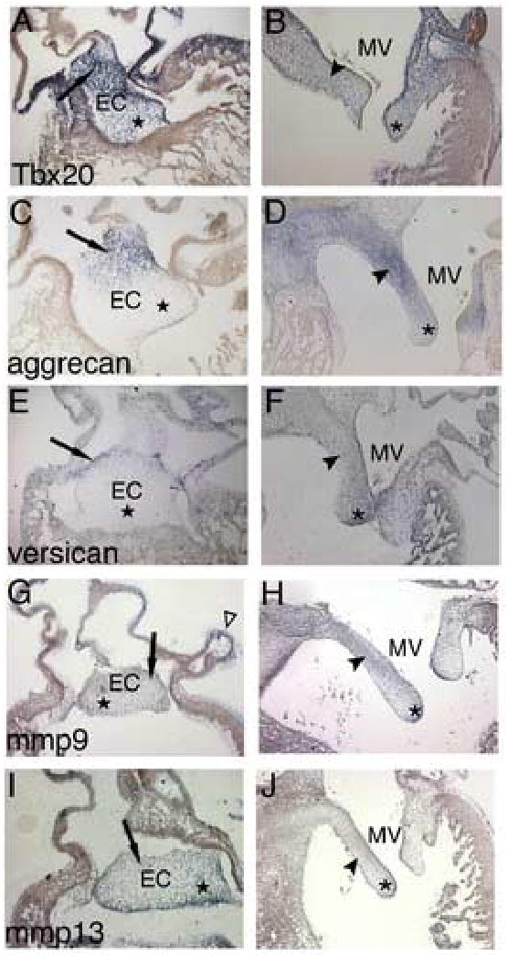
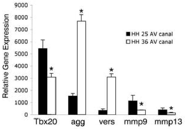
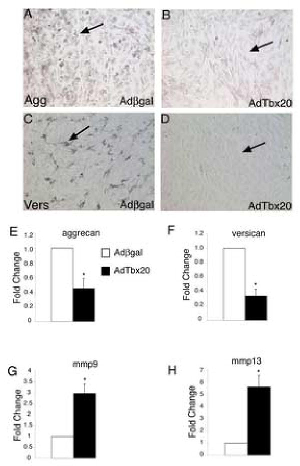
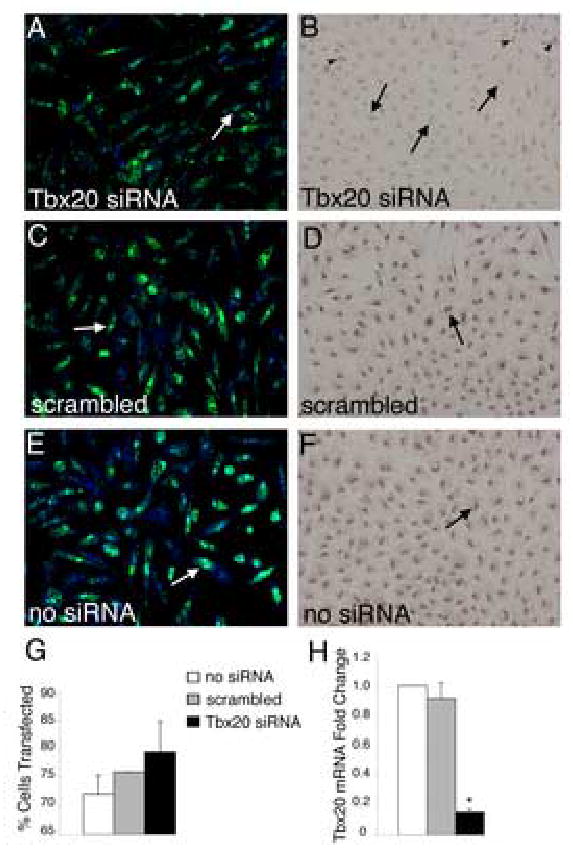
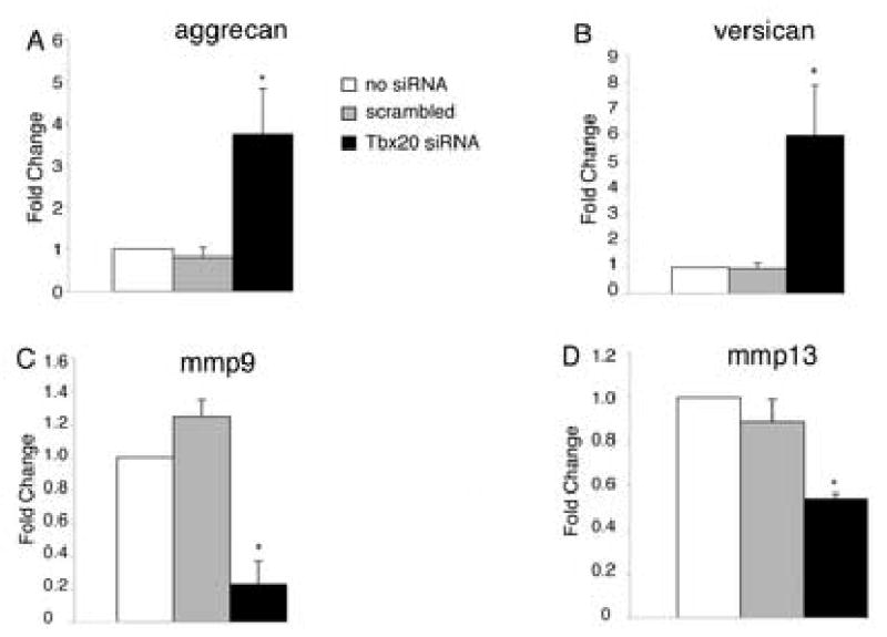
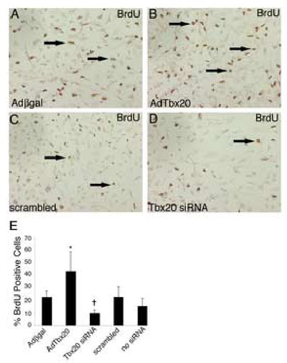
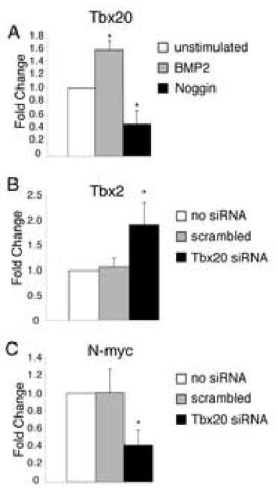
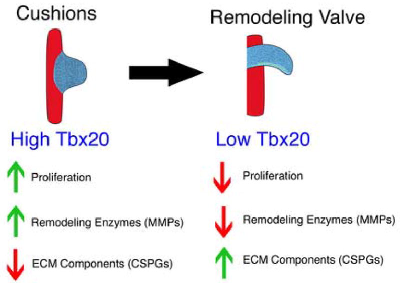
While recent work has implicated Tbx20 in myocardial maturation and proliferation, the role of Tbx20 in heart valve development remains relatively unknown. Tbx20 expression was manipulated in primary avian endocardial cells in order to elucidate its function in developing endocardial cushions. Tbx20 gain of function was achieved with a Tbx20-adenovirus, and endogenous Tbx20 expression was inhibited with Tbx20-specific siRNA in cultured endocardial cushion cells. With Tbx20 gain of function, the expression of chondroitin sulfate proteoglycans (CSPG), including aggrecan and versican, was decreased, while the expression of the matrix metalloproteinases (MMP) mmp9 and mmp13 was increased. Consistent results were observed with Tbx20 loss of function, where the expression of CSPG genes increased and MMP genes decreased. In addition, cushion mesenchyme proliferation increased with infection of a Tbx20-adenovirus and decreased with transfection of Tbx20-specfic siRNA. Furthermore, BMP2 treatment resulted in increased Tbx20 expression in endocardial cushion cells, and loss of Tbx20 led to increased Tbx2 and decreased N-myc gene expression. Taken together, these data support a role for Tbx20 in repressing extracellular matrix remodeling and promoting cell proliferation in mesenchymal valve precursor populations in endocardial cushions during embryonic development.


Figures

Figure 1
Figure 1. CSPG and MMP genes are differentially expressed in endocardial cushions and remodeling valves
Expression of Tbx20, aggrecan, versican, mmp9, and mmp13 was examined in sectioned HH stage 25 and HH stage 34 chicken hearts. In situ hybridizations show Tbx20 (A), mmp9 (G), and mmp13 (I) are expressed throughout the entire AV endocardial cushion including the subatrial region (arrow) and cushion core (star). In addition, expression of mmp9 was also detected in the subepicardial space (open arrowhead in G). In contrast, aggrecan (C) and versican (E) are expressed only in the subatrial region of the cushion (arrows). In remodeling mitral valves, Tbx20 (B) is expressed in the valve leaflet (arrowhead) and distal tips (asterisk). At HH stage 34, aggrecan (D) is expressed at high levels in the valve leaflet, but is excluded from the distal tips, while versican (F) is expressed throughout the leaflet, with increased expression at the distal tips. In remodeling valves, mmp9 (H) is expressed throughout the leaflet with concentrated expression in the atrial aspect of the leaflet and in the distal tips, while mmp13 (J) is expressed in the ventricular aspect of the leaflet and in the distal tips. EC, endocardial cushion; MV, mitral valve.
Figure 2
Figure 2. Differential expression of Tbx20, CSPG, and MMP genes during endocardial cushion and remodeling valve stages
Expression of Tbx20, aggrecan, versican, mmp9, and mmp13 was examined in isolated AV canals from HH stage 25 or HH stage 36 chicken embryos using real time RT-PCR. The expression of Tbx20, mmp9, and mmp13 was decreased at HH stage 36 compared to the expression at HH stage 25. In contrast the expression of aggrecan and versican at HH stage 36 was increased compared to the expression at HH stage 25. Values on the y-axis represent arbitrary units of fluorescence intensity for data obtained with equivalent RNA input normalized to GAPDH. These data are representative of 3 independent real time RT-PCR experiments performed in duplicate (n=3). Statistical significance of observed differences between gene expression levels at HH stage 25 and HH stage 36 is indicated by an asterisk (P<0.05), and error bars represent standard error of the mean.
Figure 3
Figure 3. Tbx20 gain of function results in decreased expression of CSPGs and increased expression of MMPs
Adenoviruses expressing β-gal (Ad β-gal) or Tbx20 (AdTbx20) were used to infect primary avian HH stage 25 endocardial cushion cells. Immunohistochemistry with antibodies specific for Aggrecan (A,B) or Versican (C,D) was used to measure aggrecan and versican protein levels following infection. Cells infected with AdTbx20 had less immunoreactivity (B,D arrows indicate lack of staining) than cells infected with Adβ-gal (A,C arrows indicate staining). These results were confirmed by real time RT-PCR. Cells infected with AdTbx20 had reduced expression of aggrecan (E) and versican (F) (n=6, P<0.01). The expression of mmp9 and mmp13 was also measured in infected cells using real time PT-PCR. In cells infected with AdTbx20, mmp9 (G) and mmp13 (H) expression was increased relative to expression in cells infected with Adβ-gal (n=6, P<0.05). Fold changes were determined by dividing experimental values by control values with control values set to 1. Asterisks indicate statistically significant differences from control values and error bars represent standard error of the mean.
Figure 4
Figure 4. Tbx20-specific siRNA transfected into primary chicken endocardial cushion cells results in decreased Tbx20 mRNA and protein expression
(A,C,E) Primary chicken endocardial cushion cells were co-transfected with siRNA and FITC-labeled fluorescent oligos (green) to visualize transfection efficiency. Cells were counterstained with TO-PRO-3 iodide (blue) to visualize the nuclei. Examples of positively transfected cells are indicated by white arrows. Transfection efficiency was determined as the number of cells with incorporation of FITC-labeled oligonucleotide/total TO-PRO-3 positive nuclei (G). These analyses demonstrated that cultures were consistently transfected at 70–80% efficiency. (B,D,F) Tbx20 protein expression was examined by immunohistochemistry with an antibody specific for Tbx20 in parallel cultures of siRNA-transfected cells. Cells transfected with Tbx20-specific siRNA (B) had significantly reduced nuclear staining (arrows indicate cells with reduced Tbx20 immunoreactivity) compared to controls (D,F, arrows indicate Tbx20 immunopositive cells). In Tbx20-specific siRNA transfected cultures (B), a subset of cells remained Tbx20 immunopositive (arrowheads). Tbx20 mRNA expression was quantified using real time RT-PCR (H). Cells transfected with Tbx20-specific siRNA had an 80% reduction in Tbx20 mRNA relative to untransfected and scrambled siRNA controls. Asterisks indicate statistically significant differences from control values and error bars represent standard error of the mean (n=3, P<0.01).
Figure 5
Figure 5. Tbx20 loss of function results in increased CSPG expression and decreased MMP expression
Primary endocardial cushion cells were transfected with Tbx20-specific or scrambled control siRNA. Cells transfected with Tbx20-specific siRNA had increased expression of aggrecan (A) and versican (B) and decreased expression of mmp9 (C) and mmp13 (D), relative to untransfected and scrambled siRNA controls as measured by real time RT-PCR. Asterisks indicate statistically significant differences from control values and error bars represent standard error of the mean (n=4, P<0.05).
Figure 6
Figure 6. Tbx20 promotes endocardial cushion cell proliferation
Primary endocardial cushion cells were infected with adenoviruses expressing β-gal (Ad β-gal) or Tbx20 (AdTbx20) for gain of function or transfected with Tbx20-specific or scrambled control siRNA for loss of function. Proliferation was measured using immunohistochemistry for BrdU incorporation. Cells infected with AdTbx20 (B) exhibit increased BrdU incorporation (arrows indicate positively stained cells) relative to cells infected with Adβ-gal (A). Cells transfected with Tbx20-specific siRNA (D) are less proliferative than cells transfected with scrambled siRNA (C). The percent of BrdU positive cells relative to total nuclei in each group is quantified (E). The asterisk represents a statistically significant difference compared to the Adβ-gal control group while the cross represents a statistically significant difference compared to the no siRNA control group (n=3, P<0.01). Error bars represent standard error of the mean.
Figure 7
Figure 7. BMP2 induces Tbx20 and Tbx20 regulates the expression of Tbx2 and N-myc in endocardial cushion cells
Endocardial cushion cells were cultured with or without the addition of soluble recombinant BMP2 or Noggin, a BMP inhibitor. Addition of BMP2 resulted in increased Tbx20 expression relative to untreated controls, while addition of Noggin attenuated that response (A), as measured by real time RT-PCR. The expression of Tbx2 and N-myc, genes associated with Tbx20 regulated proliferation, was measured in siRNA transfected endocardial cushion cells using real time RT-PCR. In cells transfected with Tbx20-specific siRNA, Tbx2 expression is increased (B) while N-myc expression is decreased relative to untransfected or scrambled siRNA controls (C). Asterisks indicate statistically significant differences from control values and error bars represent standard error of the mean (n=5, P<0.01).
Figure 8
Figure 8. Model for Tbx20 function in endocardial cushions and remodeling valves
Endocardial cushions are characterized by highly proliferative mesenchymal cells and unorganized extracellular matrix, while remodeling valves are characterized by less proliferative interstitial cells and organized extracellular matrix. These molecular and cellular characteristics are consistent with a model in which high levels of Tbx20 in endocardial cushions induce proliferation while maintaining an unremodeled, unorganized extracellular matrix. In contrast, lower levels of Tbx20 in remodeling valves allow for a less proliferative cell population and a more remodeled and organized extracellular matrix.

Similar articles

Cited by

References

    1. Akhtar S, Meek KM, James V. Ultrastructure abnormalities in proteoglycans, collagen fibrils, and elastic fibers in normal and myxomatous mitral valve chordae tendineae. Cardiovasc Pathol. 1999;8:191–201. - PubMed
    1. Arciniegas E, Neves CY, Candelle D, Parada D. Differential versican isoforms and aggrecan expression in the chicken embryo aorta. Anat Rec A Discov Mol Cell Evol Biol. 2004;279:592–600. - PubMed
    1. Armstrong EJ, Bischoff J. Heart valve development: endothelial cell signaling and differentiation. Circ Res. 2004;95:459–70. - PMC - PubMed
    1. Bailey M, Pillarisetti S, Jones P, Xiao H, Simionescu D, Vyavahare N. Involvement of matrix metalloproteinases and tenascin-C in elastin calcification. Cardiovasc Pathol. 2004;13:146–55. - PubMed
    1. Bartram U, Bartelings MM, Kramer HH, Gittenberger-de Groot AC. Congenital polyvalvular disease: a review. Pediatr Cardiol. 2001;22:93–101. - PubMed

Publication types

MeSH terms