. 2007 Mar;292(3):C1156-66.
doi: 10.1152/ajpcell.00397.2006. Epub 2006 Oct 25.

Ca2+ sparks and T tubule reorganization in dedifferentiating adult mouse skeletal muscle fibers

Affiliations

Ca2+ sparks and T tubule reorganization in dedifferentiating adult mouse skeletal muscle fibers

Lisa D Brown et al. Am J Physiol Cell Physiol. 2007 Mar.

Abstract

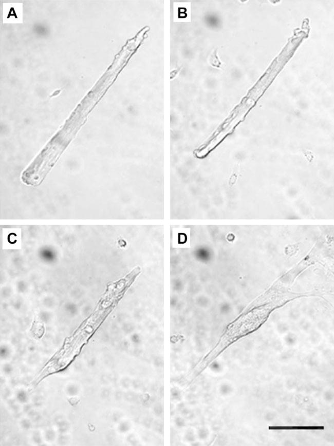
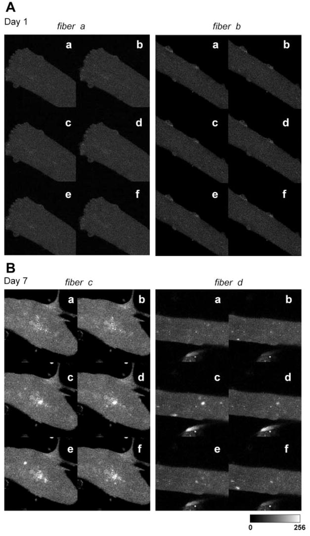
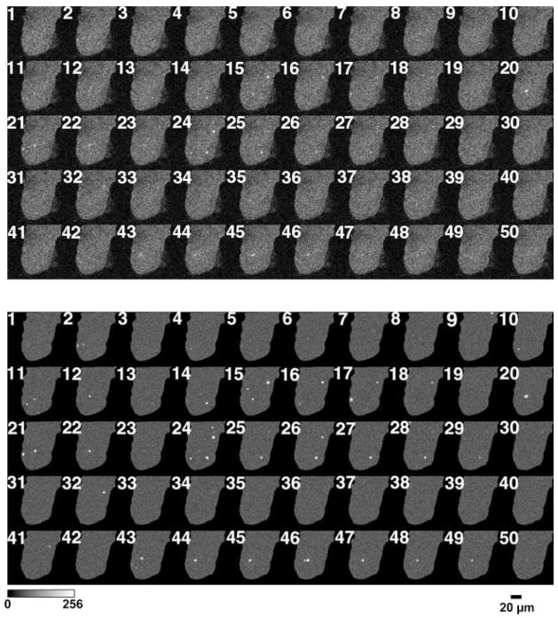
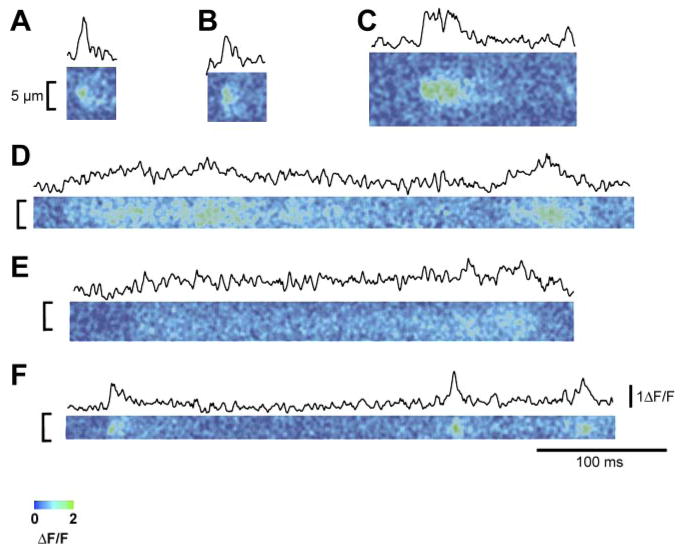
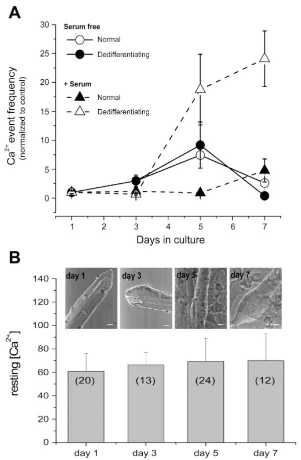
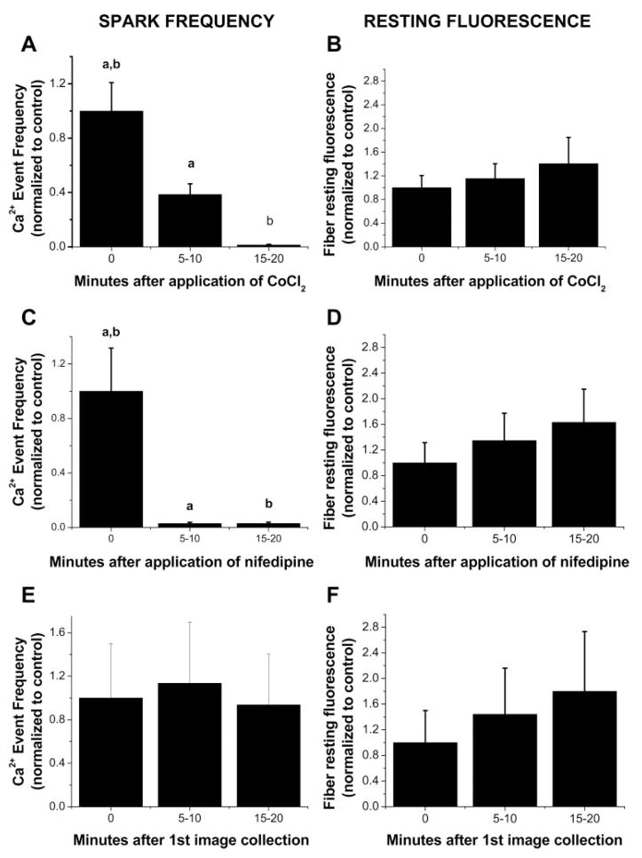
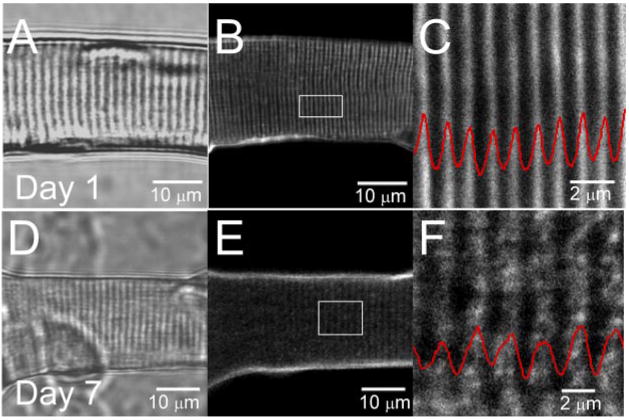
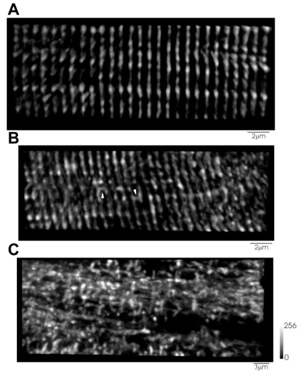
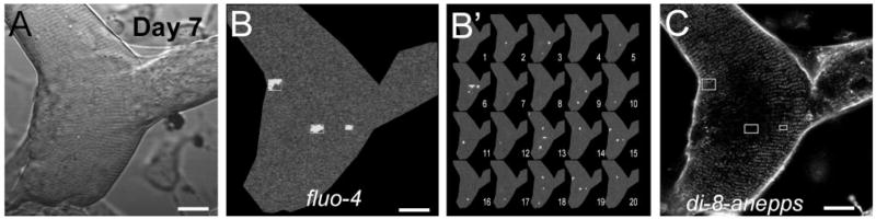
Ca(+) sparks are rare in healthy adult mammalian skeletal muscle but may appear when adult fiber integrity is compromised, and occur in embryonic muscle but decline as the animal develops. Here we used cultured adult mouse flexor digitorum brevis muscle fibers to monitor occurrence of Ca(2+) sparks during maintenance of adult fiber morphology and during eventual fiber morphological dedifferentiation after various times in culture. Fibers cultured for up to 3 days retain normal morphology and striated appearance. Ca(2+) sparks were rare in these fibers. At 5-7 days in culture, many of the original muscle fibers exhibit sprouting and loss of striations, as well as the occurrence of spontaneous Ca(2+) sparks. The average rate of occurrence of Ca(2+) sparks is >10-fold higher after 5-7 days in culture than in days 1-3. With the use of fibers cultured for 7 days, application of the Ca(2+) channel blockers Co(2+) or nifedipine almost completely suppressed the occurrence of Ca(2+) sparks, as previously shown in embryonic fibers, suggesting that Ca(2+) sparks may be generated by similar mechanisms in dedifferentiating cultured adult fibers and in embryonic fibers before final differentiation. The sarcomeric disruption observed under transmitted light microscopy in dedifferentiating fibers was accompanied by morphological changes in the transverse (T) tubular system, as observed by fluorescence confocal imaging of both an extracellular marker dye and membrane staining dyes. Changes in T tubule morphology coincided with the appearance of Ca(2+) sparks, suggesting that Ca(2+) sparks may either be a signal for, or the result of, disruption of DHPR-ryanodine receptor 1 coupling.


Figures

Fig. 1
Fig. 1
A single representative isolated adult flexor digitorum brevis (FDB) muscle fiber cultured on a laminin-coated cover slip in serum-containing media. Transmitted light images of the same fiber were taken 1 day (A), 3 days (B), 5 days (C), and 7 days (D) after plating. The images show the gradual transition from the adult fiber appearance to a myotube-like morphology, including branches and fine projections. The culture was returned to the tissue culture incubator for 2-day intervals between acquisitions of successive images. Scale bar = 0.1 mm.
Fig. 2
Fig. 2
Local Ca2+ sparks events in single muscle fibers after 1 day (A) and after 7 days (B) in culture. Six representative images of 2 different fibers are shown for each condition (a–f). Note the appearance of Ca2+ events in the fibers cultured for 7 days.
Fig. 3
Fig. 3
Ca2+ sparks appear in both the periphery and the center of dedifferentiating muscle fibers. A: 50 sequential full-frame fluorescence images of a muscle fiber loaded with fluo 4 after being maintained in a serum-containing culture for 7 days. B: events selected from A as being within the selection criteria of contiguous pixels >2 SD above the mean of the same pixel, with at least one pixel >3 SD above the mean. Each selected event is shown within an enclosing box in the image. Successive images were recorded at 3-s intervals.
Fig. 4
Fig. 4
ΔF/F (where F is fluorescence) line scan images from intact dedifferentiating adult mouse muscle fibers. Above each ΔF/F image is the mean temporal profile centered at the peak ΔF/F. These records represent a sampling of the various different time courses of local Ca2+ release events recorded from dedifferentiating fibers using line scan imaging. A, B, and F: line scan images and time courses of both rapidly rising and decaying fluorescence signals. Note that these signals are quite similar to Ca2+ sparks observed in frog and mammalian myofibers. C, D, and E: line scan images and time courses of slower rising and decaying events. Note that these records are similar to Ca2+ signals as seen in developing mammalian myofibers.
Fig. 5
Fig. 5
Frequency of Ca2+ events in both normal and dedifferentiated adult FDB fibers and resting Ca2+ concentration ([Ca2+]) after various times in culture. A: fibers were cultured in either serum-containing media or serum-free media. Frequency of Ca2+ event fibers increases as fiber dedifferentiation increases. In serum-containing media, the no. of fibers included with normal appearance was 15, 20, 9, and 3 and 0, 0, 11, and 5 with dedifferentiated appearance at the successive time points, and in serum-free media was 25, 16, 19, and 10 with normal appearance and 0, 2, 5, and 7 with dedifferentiated appearance at the successive time points. Statistical significance was determined using a 2-way ANOVA test. Significance (P < 0.05) was found between the dedifferentiating fibers in serum-containing media and all the other groups at both days 5 and 7. B: intracellular resting [Ca2+] during fiber dedifferentiation. Bar graph summarizing mean resting [Ca2+] evaluated by ratiometric photometry in indo 1-loaded FDB fibers cultured in serum-containing media at four different stages (days 1, , , and 7) after fiber plating. Transmitted light images at top of each column shows the typical appearance of FDB fibers for each stage. Scale bar = 10 μm; nos. in parentheses represent sample size.
Fig. 6
Fig. 6
Frequency of Ca2+ events in dedifferentiated adult FDB fibers decreases with addition of Ca2+ channel blockers. All fibers were cultured for 6–7 days in CoCl2-free, nifedipine-free, serum-containing medium before each experiment. A: fibers cultured in serum-containing media were bathed in Ringer solution and imaged both before and after the addition of 5 mM CoCl2 as indicated. B: time course of resting fiber fluorescence in the same fibers both before and after the addition of CoCl2. C: fibers cultured in serum-containing media were bathed in Ringer solution and imaged both before and after the addition of 5 μM nifedipine as indicated. D: time course of resting fiber fluorescence in the same fibers both before and after the addition of nifedipine. E: fibers cultured in serum-containing media were bathed in Ringer solution and imaged for spark activity without Ca2+ channel blocker treatment. F: time course of resting fiber fluorescence in the same fibers without Ca2+ channel blocker treatment; n = 21 in A and B, n = 10 in C and D, and n = 10 in E and F. Statistical significance was determined using a 1-way ANOVA test (P < 0.05). Groups with the same letter exhibit significant difference from each other.
Fig. 7
Fig. 7
Transmitted light (A–C) and sulforhodamine B (A′–C′) images of fibers after 1 and 7 days in culture. After 1 day in culture, both transmitted light (A) and sulforhodamine-stained (A′) images showed an orderly pattern of both striations and transverse (T) tubule structure. By day 7, dedifferentiating fibers exhibit various stages of disruption of T tubule structure after staining with sulforhodamine B (B′ and C′). Longitudinally oriented T tubules are apparent in C′.
Fig. 8
Fig. 8
T tubule structure reorganization during fiber dedifferentiation. Transmitted light images (average of 8 frames) of FDB fibers cultured in serum-containing media after 1 (A) and 7 (B) days in culture. B and E: laser confocal images (average of 16 frames) of the same fibers stained with pyridinium, 4-[2-(6-(dioctylamino)-2-naphthalenyl) ethenyl]-1-(3-sulfopro-pyl)-, inner salt (di-8-ANEPPS) from a single z-axis plane. C and F: enlarged views of the outlined regions of the fibers shown in B and E, respectively. Red traces, average intensity profile, plotted as a function of the longitudinal position, of area enclosed within white rectangles in B and E. After 7 days in culture, T tubular structure was disorganized and characterized by an apparent reduction in the T tubule spacing.
Fig. 9
Fig. 9
T tubule structure in fibers after culture for 1 and 7 days: FM4–64 staining. A: FM4–64 staining of T tubules shows a normal transverse striated pattern after 1 day in culture. B and C: after 7 days in culture, T tubules show an alteration in the transverse striated pattern, with either an occasional longitudinal T tubule (arrowhead; B) or (C) a complete loss of the transverse striated pattern with areas of the fiber void of T tubules (C).
Fig. 10
Fig. 10
T tubule reorganization and Ca2+ sparks in a di-8-ANEPPS-stained and fluo 4-loaded day 7 dedifferentiated fiber. A series of images of fluorescence of fluo-4 (50 frames) and fluorescence of di-8-ANEPPS (average of 8 images) was obtained in a sequential mode scanning in the same fiber. A: transmitted light image. B: representative single full x-y frame image showing Ca2+ sparks appearing in both the periphery and the center of day 7 dedifferentiating muscle fiber. Each selected event is shown within an enclosing box in the image. B′: 20 sequential x-y mode images of fiber shown in B. Events were selected as in Fig. 3. C: image of fluorescence of di-8-ANEPPS, showing reorganized T tubules. For comparison, enclosing boxes showing events in B, were redrawn here. Note that Ca2+ sparks usually occur in central areas, where T tubule disruption is more dramatic. Scale bars = 10 μm.

References

    1. Block BA, Imagawa T, Campbell KP, Franzini-Armstrong C. Structural evidence for direct interaction between the molecular components of the transverse tubule-sarcoplasmic reticulum junction in skeletal muscle. J Cell Biol. 1988;107:2587–2600. - PMC - PubMed
    1. Brown LD, Schneider MF. Delayed differentiation and retention of properties in dissociated adult skeletal muscle fibers in vitro. In Vitro Cell Dev Biol-Animal. 2002;38:411–422. - PubMed
    1. Chen YW, Zhao P, Borup R, Hoffmann EP. Expression profiling in the muscular dystrophies: identification of novel aspects of molecular patho-physiology. J Cell Biol. 2000;151:1321–1336. - PMC - PubMed
    1. Cheng H, Lederer WJ, Cannell MB. Calcium sparks: elementary events underlying excitation-contraction coupling in heart muscle. Science. 1993;262:740–744. - PubMed
    1. Cheng H, Song LS, Shirokova N, Gonzalez A, Lakatta EG, Rios E, Stern MD. Amplitude distribution of calcium sparks in confocal images: theory and studies with an automatic detection method. Biophys J. 1999;76:606–617. - PMC - PubMed

LinkOut - more resources