Sympathetic sprouting near sensory neurons after nerve injury occurs preferentially on spontaneously active cells and is reduced by early nerve block
- PMID: 17065247
- PMCID: PMC1774587
- DOI: 10.1152/jn.00899.2006
Sympathetic sprouting near sensory neurons after nerve injury occurs preferentially on spontaneously active cells and is reduced by early nerve block
Abstract
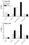
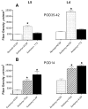
Some chronic pain conditions are maintained or enhanced by sympathetic activity. In animal models of pathological pain, abnormal sprouting of sympathetic fibers around large- and medium-sized sensory neurons is observed in dorsal root ganglia (DRGs). Large- and medium-sized cells are also more likely to be spontaneously active, suggesting that sprouting may be related to neuron activity. We previously showed that sprouting could be reduced by systemic or locally applied lidocaine. In the complete sciatic nerve transection model in rats, spontaneous activity initially originates in the injury site; later, the DRGs become the major source of spontaneous activity. In this study, spontaneous activity reaching the DRG soma was reduced by early nerve blockade (local perfusion of the transected nerve with TTX for the 1st 7 days after injury). This significantly reduced sympathetic sprouting. Conversely, increasing spontaneous activity by local nerve perfusion with K(+) channel blockers increased sprouting. The hyperexcitability and spontaneous activity of DRG neurons observed in this model were also significantly reduced by early nerve blockade. These effects of early nerve blockade on sprouting, excitability, and spontaneous activity were all observed 4-5 wk after the end of early nerve blockade, indicating that the early period of spontaneous activity in the injured nerve is critical for establishing the more long-lasting pathologies observed in the DRG. Individual spontaneously active neurons, labeled with fluorescent dye, were five to six times more likely than quiescent cells to be co-localized with sympathetic fibers, suggesting a highly localized correlation of activity and sprouting.
Figures


Similar articles
-
Local knockdown of the NaV1.6 sodium channel reduces pain behaviors, sensory neuron excitability, and sympathetic sprouting in rat models of neuropathic pain.Neuroscience. 2015 Apr 16;291:317-30. doi: 10.1016/j.neuroscience.2015.02.010. Epub 2015 Feb 14. Neuroscience. 2015. PMID: 25686526 Free PMC article.
-
Absence of an association between axotomy-induced changes in sodium currents and excitability in DRG neurons from the adult rat.Pain. 2004 Jun;109(3):471-480. doi: 10.1016/j.pain.2004.02.024. Pain. 2004. PMID: 15157708
-
Early blockade of injured primary sensory afferents reduces glial cell activation in two rat neuropathic pain models.Neuroscience. 2009 Jun 2;160(4):847-57. doi: 10.1016/j.neuroscience.2009.03.016. Epub 2009 Mar 19. Neuroscience. 2009. PMID: 19303429 Free PMC article.
-
Recent evidence for activity-dependent initiation of sympathetic sprouting and neuropathic pain.Sheng Li Xue Bao. 2008 Oct 25;60(5):617-27. Sheng Li Xue Bao. 2008. PMID: 18958370 Review.
-
Contribution of the spared primary afferent neurons to the pathomechanisms of neuropathic pain.Mol Neurobiol. 2002 Aug;26(1):57-67. doi: 10.1385/MN:26:1:057. Mol Neurobiol. 2002. PMID: 12392056 Review.
Cited by
-
Governing role of primary afferent drive in increased excitation of spinal nociceptive neurons in a model of sciatic neuropathy.Exp Neurol. 2008 Dec;214(2):219-28. doi: 10.1016/j.expneurol.2008.08.003. Epub 2008 Aug 16. Exp Neurol. 2008. PMID: 18773893 Free PMC article.
-
Local knockdown of the NaV1.6 sodium channel reduces pain behaviors, sensory neuron excitability, and sympathetic sprouting in rat models of neuropathic pain.Neuroscience. 2015 Apr 16;291:317-30. doi: 10.1016/j.neuroscience.2015.02.010. Epub 2015 Feb 14. Neuroscience. 2015. PMID: 25686526 Free PMC article.
-
Efficacy of the lumbar sympathetic ganglion block in lower limb pain and its application prospects during the perioperative period.Ibrain. 2022 Oct 9;8(4):442-452. doi: 10.1002/ibra.12069. eCollection 2022 Winter. Ibrain. 2022. PMID: 37786587 Free PMC article. Review.
-
In Pursuit of an Allosteric Human Tropomyosin Kinase A (hTrkA) Inhibitor for Chronic Pain.ACS Med Chem Lett. 2021 Oct 25;12(11):1847-1852. doi: 10.1021/acsmedchemlett.1c00483. eCollection 2021 Nov 11. ACS Med Chem Lett. 2021. PMID: 34795875 Free PMC article.
-
Upregulation of the sodium channel NaVβ4 subunit and its contributions to mechanical hypersensitivity and neuronal hyperexcitability in a rat model of radicular pain induced by local dorsal root ganglion inflammation.Pain. 2016 Apr;157(4):879-891. doi: 10.1097/j.pain.0000000000000453. Pain. 2016. PMID: 26785322 Free PMC article.
References
-
- Abdulla FA, Moran TD, Balasubramanyan S, Smith PA. Effects and consequences of nerve injury on the electrical properties of sensory neurons. 2003;81:663–682. - PubMed
-
- Babbedge RC, Soper AJ, Gentry CT, Hood VC, Campbell EA, Urban L. In vitro characterization of a peripheral afferent pathway of the rat after chronic sciatic nerve section. J Neurophysiol. 1996;76:3169–3177. - PubMed
-
- Boucher TJ, Okuse K, Bennett DL, Munson JB, Wood JN, McMahon SB. Potent analgesic effects of GDNF in neuropathic pain states. Science. 2000;290:124–127. - PubMed
Publication types
MeSH terms
Substances
Grants and funding
LinkOut - more resources
Full Text Sources
Other Literature Sources
Medical

