. 2006 Nov;47(11):4687-99.
doi: 10.1167/iovs.05-1369.

Effectiveness of hyperopic defocus, minimal defocus, or myopic defocus in competition with a myopiagenic stimulus in tree shrew eyes

Affiliations

Effectiveness of hyperopic defocus, minimal defocus, or myopic defocus in competition with a myopiagenic stimulus in tree shrew eyes

Thomas T Norton et al. Invest Ophthalmol Vis Sci. 2006 Nov.

Abstract

Purpose: To examine the ability of hyperopic defocus, minimal defocus, and myopic defocus to compete against a myopiagenic -5-D lens in juvenile tree shrew eyes.

Methods: Juvenile tree shrews (n > or = 5 per group), on a 14-hour lights-on/10-hour lights-off schedule, wore a monocular -5-D lens (a myopiagenic stimulus) over the right eye in their home cages for more than 23 hours per day for 11 days. For 45 minutes each day, the animals were restrained so that all visual stimuli were >1 m away. While viewing distance was controlled, the -5-D lens was removed and another lens was substituted with one of the following spherical powers: -5 D, -3 D (hyperopic defocus); plano (minimal defocus); or +3, +4, +5, +6, or +10 D (myopic defocus). Daily noncycloplegic autorefractor measures were made on most animals. After 11 days of treatment, cycloplegic refractive state and axial component dimensions were measured.

Results: Eyes with the substituted -5- or -3-D-lens developed significant myopia (mean +/- SEM, -4.7 +/- 0.3 and -3.1 +/- 0.1 D, respectively) and appropriate vitreous chamber elongation. All animals with the substituted plano lens (minimal defocus) during the 45-minute period showed no axial elongation or myopia (the plano lens competed effectively against the -5-D lens). Variable results were found among animals that wore a plus lens (myopic defocus). In 11 of 20 eyes, a +3-, +4-, or +5-D lens competed effectively against the -5-D lens (treated eye <1.5 D myopic relative to its fellow control eye). In the other eyes (9/20) myopic defocus was ineffective in blocking compensation; the treated eye became more than 2.5 D myopic relative to the control eye. The +6- and +10-D substituted lenses were ineffective in blocking compensation in all cases.

Conclusions: When viewing distance was limited to objects >1 m away, viewing through a plano lens for 45 minutes (minimal defocus) consistently prevented the development of axial elongation and myopia in response to a myopiagenic -5-D lens. Myopic defocus prevented compensation in some but not all animals. Thus, myopic defocus is encoded by at least some tree shrew retinas as being different from hyperopic defocus, and myopic defocus can sometimes counteract the myopiagenic effect of the -5-D lens (hyperopic defocus). However, it appears that minimal defocus is a more consistent, strong antidote to a myopiagenic stimulus in this mammal closely related to primates.


Figures

Figure 1
Figure 1
(A) Difference in cycloplegic refraction (treated minus control eye), measured with no lenses present, after 11 days of lens treatment in the eight groups of tree shrews. Error bars are SEM. The power of the CVD lens worn 45 min/d is indicated on the abscissa. Differences for individual animals are represented by the filled and open circles. Filled symbols indicate animals in which the plus lens blocked compensation to the −5-D lens; the treated eye was myopic, relative to the control eye, by less than 1.5 D. (B) Mean and individual differences in vitreous chamber depth. Probabilities are the results of one-tailed t-tests that examined whether the treated eyes in each group were significantly myopic, or elongated, compared with the control eyes.
Figure 2
Figure 2
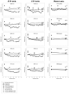
Noncycloplegic and cycloplegic autorefractor measures in the animals that wore a −5-D lens in the home cage and that wore (left column)a −5-D CVD lens, (center column) a −3-D CVD lens, or (right column) a plano CVD lens. Square symbols: measures made with the −5-D lens in place. Filled and open triangles and the gray square: measures made with atropine cycloplegia. For the −3-D and plano CVD lens groups the gray diamond indicates cycloplegic refractive measures made while the animals wore the CVD lens. The cycloplegic measures were made 1 to 2 hours after the final noncycloplegic measures and are displaced along the abscissa for clarity. The horizontal line at 4 D represents estimated emmetropia in tree shrews when measured with the autorefractor.
Figure 3
Figure 3
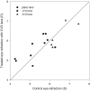
Comparison of cycloplegic control-eye refractive measures versus treated-eye measures while the treated eyes wore the CVD lens. The solid line indicates equal refraction in each eye. The control eye refraction varied between 4.2 and 7.5 D, and the treated eyes, while wearing the CVD lens, were all within 0.9 D of their control eye after 11 days of treatment. This is within the estimated depth of focus of the tree shrew eye (±1.0−1.5 D).,
Figure 4
Figure 4
Distribution of cycloplegic refractive differences (treated minus control eye) in the groups of animals that wore a plus CVD lens. Eyes that became more than 2.5 D myopic, relative to their fellow control eye, lie to the left of the dashed line Eyes that developed less than 1.5 D of myopia lie to the right of the dashed line The power of the CVD lens is indicated in the key.
Figure 5
Figure 5
Autorefractor measures of the animals that wore a plus lens while viewing distance was controlled. Within a group, animals are placed vertically in the order from the least amount of compensation (CVD lens competed most effectively) to the most (CVD lens least effective). Horizontal lines: separate animals in the group in which compensation was blocked (above the line) from those in which it was not blocked (below the line) as indicated in Figures 1 and 4. For the +5-D CVD lens group, the entire left column is above the line. Other symbols are as in Figure 2.
Figure 6
Figure 6
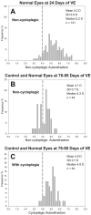
Frequency distribution of spherical-equivalent refraction for normal and control tree shrew eyes. (A) Noncycloplegic measures of normal eyes at 24 days of VE (equivalent to treatment day 1). (B) Noncycloplegic measures of control and normal (right and left averaged) eyes at 75 to 96 days of VE. (C) The same eyes as in (B), measured under atropine cycloplegia. Dashed vertical line: at +4 D represents estimated emmetropia. The bin width is 0.2 D.
Figure 7
Figure 7
Calculated amount of defocus (without regard to sign) experienced by the treated eyes of the plus-lens CVD animals on day 1 of treatment while wearing the −5-D lens and the CVD lens, based on noncycloplegic refractions made with the lenses in place on treatment day 1. In this figure, it is assumed that a +4-D autorefractor reading actually is emmetropia and that there is no change in accommodation from the values measured with the autorefractor. Filled symbols: eyes in which the plus CVD lens blocked compensation to the −5-D lens. Open symbols: eyes that, by treatment day 12, had compensated for the −5-D lens. Solid line: equal defocus in both conditions measured at optical infinity. Data were not obtained for one animal in the +10-D CVD-lens group. DNB, did not block. The circled data point is the animal that developed significant hyperopia.

References

    1. Norton TT, McBrien NA. Normal development of refractive state and ocular component dimensions in the tree shrew (Tupaia belangeri) Vision Res. 1992;32:833–842. - PubMed
    1. Pickett-Seltner RL, Sivak JG, Paternak JJ. Experimentally induced myopia in chicks: morphometric and biochemical analysis during the first 14 days after hatching. Vision Res. 1988;28:323–328. - PubMed
    1. Smith EL, III, Hung LF. The role of optical defocus in regulating refractive development in infant monkeys. Vision Res. 1999;39:1415–1435. - PubMed
    1. Bradley DV, Fernandes A, Lynn M, Tigges M, Boothe RG. Emmetropization in the rhesus monkey (Macaca mulatta): birth to young adulthood. Invest Ophthalmol Vis Sci. 1999;40:214–229. - PubMed
    1. Cook RC, Glasscock RE. Refractive and ocular findings in the newborn. Am J Ophthalmol. 1951;34:1407–1413. - PubMed

Publication types