. 2007 Jan;18(1):1-13.
doi: 10.1091/mbc.e06-08-0753. Epub 2006 Oct 25.

The NHR1 domain of Neuralized binds Delta and mediates Delta trafficking and Notch signaling

Affiliations

The NHR1 domain of Neuralized binds Delta and mediates Delta trafficking and Notch signaling

Cosimo Commisso et al. Mol Biol Cell. 2007 Jan.

Abstract

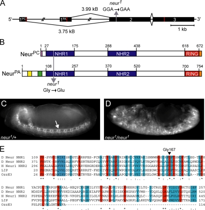
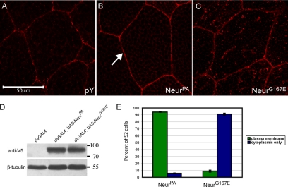
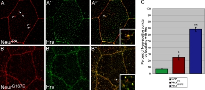
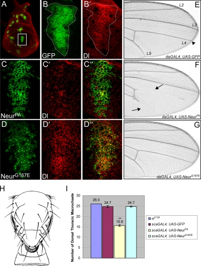
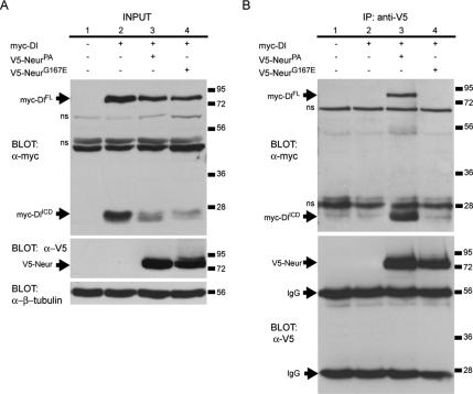
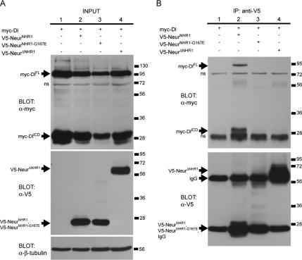
Notch signaling, which is crucial to metazoan development, requires endocytosis of Notch ligands, such as Delta and Serrate. Neuralized is a plasma membrane-associated ubiquitin ligase that is required for neural development and Delta internalization. Neuralized is comprised of three domains that include a C-terminal RING domain and two neuralized homology repeat (NHR) domains. All three domains are conserved between organisms, suggesting that these regions of Neuralized are functionally important. Although the Neuralized RING domain has been shown to be required for Delta ubiquitination, the function of the NHR domains remains elusive. Here we show that neuralized, a well-characterized neurogenic allele, exhibits a mutation in a conserved residue of the NHR1 domain that results in mislocalization of Neuralized and defects in Delta binding and internalization. Furthermore, we describe a novel isoform of Neuralized and show that it is recruited to the plasma membrane by Delta and that this is mediated by the NHR1 domain. Finally, we show that the NHR1 domain of Neuralized is both necessary and sufficient to bind Delta. Altogether, our data demonstrate that NHR domains can function in facilitating protein-protein interactions and in the case of Neuralized, mediate binding to its ubiquitination target, Delta.


Figures

Figure 1.
Figure 1.
The neurogenic allele neur1 contains a mutation resulting in the subsitution of a conserved Gly residue of NHR1 with a Glu. (A) The neur locus produces two transcripts with unique first exons. The second exon mutation present in the neur1 allele is indicated with a gray arrow. Red lines indicate the ATG start sites and translational STOP codons. (B) The resulting protein isoforms differ at their N-termini. NHR domains (blue) and the RING domain (red) are present in both isoforms. NeurPA unique regions include the glutamine/histidine-rich region (yellow) and the lysine/arginine-rich region (green). NeurPC exhibits an eight amino acid unique region (pink). For ectopic expression V5 epitope tags are located at the carboxyl termini (orange). The G167E mutation present in neur1 is indicated with a gray arrow (residue numbering is in reference to NeurPA). (C and D) Embryos from neur1/TM3, Sb were collected and stained with FITC-conjugated anti-HRP, which labels the CNS. neur1 heterozygous embryos (C) and neur1/neur1 homozygous mutant embryos (D) are shown. The CNS of heterozygous embryos is indistinguishable from wild-type and is indicated by the arrow in C. neur1/neur1 mutant embryos exhibit a neurogenic phenotype consisting of excess neural tissue at the expense of epidermis (D). (E) A multiple sequence alignment of NHR domains reveals highly conserved residues (red) and residues with semiconserved substitutions (blue). Gly167, the amino acid residue affected in neur1, is one of the highly conserved residues and is indicated by the arrowhead. Sequence alignment was generated using ClustalW (http://www.ebi.ac.uk/clustalw/). Representative NHR domains are from Drosophila Neuralized (D Neur, GenPept NP_476652, residue numbering is in reference to NeurPA), human Neuralized-1 (H Neur1, GenPept NP_004201), mouse LINCR (LIP, GenPept NP_700457), and mouse OzzE3 (GenPept Q9D0S4).
Figure 2.
Figure 2.
NeurG167E exhibits increased localization to cytoplasmic puncta compared with wild-type NeurPA. (A) Salivary gland cells stained with anti-phosphotyrosine, a plasma membrane marker (shown in red). (B and C) Staining for V5-tagged NeurPA (B) or NeurG167E (C) in larval salivary glands (shown in red). Transgenes are expressed using scaGAL4. As expected, NeurPA exhibits predominantly plasma membrane localization as indicated by the arrow in B. NeurG167E is present in many more cytoplasmic puncta than wild type (C). (D) Western analysis of adult lysates of the genotype indicated. Neur proteins are detected using anti-V5. β-tubulin is shown as a loading control. The neur transgenes are expressed at comparable levels. (E) Quantification of subcellular localization in S2 cells. NeurPA is localized to the plasma membrane in >90% of cells. In contrast, NeurG167E is localized to the plasma membrane in <10% of cells and is predominantly cytoplasmic (>90%). For each construct, analysis was done in triplicate (n = 3) with a sample size of 100. Error bars, SE.
Figure 3.
Figure 3.
The G167E mutation increases Neur localization to Hrs-containing endosomes in vivo. (A and B) Staining for V5-tagged NeurPA (A) or NeurG167E (B) is shown in red, whereas staining for the endosomal marker Hrs (A′ and B′) is shown in green. Overlays are shown in A″ and B″ and colocalization is shown in yellow. Insets are digital magnifications of regions within each cell as indicated by the asterisks. V5-NeurPA, which is predominantly localized to the plasma membrane, exhibits a low level of cytoplasmic staining as indicated by the arrowheads in A. Some of these cytoplasmic puncta colocalize with Hrs as shown in A″. V5-NeurG167E, which exhibits many more cytoplasmic puncta than wild-type (B), also colocalizes with Hrs to a much higher degree (B″). (C) Quantification of colocalization between Hrs and GFP, NeurPA, or NeurG167E. As a control, the extent of colocalization between GFP and Hrs was determined. This low level of colocalization (∼7%) is considered baseline. NeurPA colocalizes with Hrs to a much higher extent than baseline (∼24%) and is statistically significant, indicating the bona fide presence of wild-type Neur in Hrs endosomes. NeurG167E exhibits a much higher degree of colocalization with Hrs than wild type (just below 70%). This increase is statistically significant compared with wild type. For each genotype, four cells from different salivary glands were sectioned throughout and vesicles counted (n = 4). Error bars, SE. The asterisks indicate statistical significance (*p < 0.05, **p < 0.001).
Figure 4.
Figure 4.
A mutation in NHR1 prevents NeurG167E from increasing Dl internalization or disrupting N-dependent tissue development. (A) Expression of UAS-GFP in the wing disc (shown in green) using the scaGAL4 enhancer trap labels proneural regions. Dl staining (shown in red) is used to visualize the disc. (B and B′) A higher magnification of the presumptive wing vein tissue (white box in A). scaGAL4 expression boundaries are shown by GFP expression (shown in green, B). Dl staining is in red (B′). In this tissue, Dl is localized to the plasma membrane and in cytoplasmic puncta (B). (C and D). Same region as outlined in B when either V5-NeurPA (C) or V5-NeurG167E (D) is expressed (shown in green). Dl staining is shown in red (C′ and D′). Overlays are shown in C″ and D″. Colocalization is shown in yellow. All images were obtained from the same plane in the apical part of the wing disc. NeurPA is localized to the plasma membrane (C) and increases Dl internalization resulting in reduced levels of Dl at the plasma membrane (C′). NeurPA and Dl show little colocalization (C″). It should be noted that in other parts of the wing disc, Dl did maintain some plasma membrane localization, but was still reduced. In contrast, NeurG167E is predominantly localized to cytoplasmic puncta (D) and does not reduce plasma membrane levels of Dl (D′). NeurG167E and Dl exhibit colocalization in cytoplasmic puncta (D″). (E–G) Distal regions of adult wings expressing GFP (E), NeurPA (F), or NeurG167E (G) using the ubiquitous driver daGAL4. In wild-type wings, veins extend to the wing margin (arrowhead in E). When NeurPA is expressed, wing veins are truncated (arrows in F). In contrast, wing veins are unaffected when NeurG167E is expressed (G). (H) The adult dorsal thorax exhibits 26 large bristles, known as macrochaetes (adapted from Ferris, 1950). (I) Quantification of the number of macrocheates in flies expressing GFP, NeurPA, or NeurG167E compared with wild-type (w1118). Expression of NeurPA affects sense organ determination and results in reduced numbers of dorsal thoracic macrocheates compared with wild type and GFP controls. In contrast, NeurG167E does not alter the number of macrocheates compared with controls. Ten flies for each genotype were analyzed (n = 10). Error bars, SE. The asterisks indicate statistical significance (**p < 0.001).
Figure 5.
Figure 5.
The G167E mutation in the Neur NHR1 domain disrupts binding to Dl. (A) Western blots showing experimental input. Top, anti-myc labels Dl full-length (myc-DlFL) and the Dl intracellular domain (myc-DlICD) in lysates from transfected S2 cells (lanes 2–4). Lysate from untransfected cells is analyzed in lane 1. Middle, anti-V5 labels experimental input from lysates containing V5-NeurPA or V5-NeurG167E. Bottom, β-tubulin is used as a loading control. Nonspecific bands are indicated (ns). (B) Western analysis of coimmunoprecipitation assays. Top, myc-DlFL and myc-DlICD are only coimmunoprecipitated with wild-type NeurPA (lane 3). Dl does not coimmunoprecipitate in the absence of any V5-tagged Neur protein (lane 2) and does not coimmunoprecipitate with NeurG167E (lane 4). Nonspecific bands are indicated (ns). Bottom, V5-NeurPA (lane 3) and V5-NeurG167E (lane 4) are immunoprecipitated at similar levels. Anti-V5 also labels the immunoglobulin heavy and light chains (IgG) as indicated.
Figure 6.
Figure 6.
The Neur isoforms are differentially localized in S2 cells but not in vivo. (A and B) V5-tagged NeurPA (A) and NeurPC (B) proteins were constitutively expressed in S2 cells. Cells were stained with anti-V5 (shown in red, A and B) and the nuclear marker DAPI (shown in blue, A′ and B′). Channel overlays are shown in A″ and B″. Note that NeurPA exhibits predominantly plasma membrane localization, indicated by the arrow in A. In contrast, NeurPC exhibits cytoplasmic localization (B). (C and D) V5-tagged NeurPA (C) and NeurPC (D) transgenes were expressed in proneural regions using scaGAL4. Third larval instar wing discs were stained with anti-V5 (shown in red). Embryonic localization of the Neur isoforms was also analyzed (C and D, insets). In both cases, the Neur isoforms exhibit predominantly plasma membrane localization.
Figure 7.
Figure 7.
Dl mediates plasma membrane recruitment of NeurPC via NHR1. (A) Various V5-tagged Neur truncations used in this study. (B, C, and D). S2 cells were cotransfected with the Neur construct indicated (shown in red, B′, C′, and D′) and myc-tagged Dl (shown in green, B, C, and D). Overlays are shown in B″, C″, and D″ and colocalization is indicated in yellow. Dl is able to recruit NeurPC to the membrane as shown by the arrow in B′. NHR1 is sufficient for plasma membrane recruitment by Dl as indicated by the arrow in C′. The cell to the right in C′ (indicated by the arrowhead) is singly transfected with NeurNHR1 and does not exhibit plasma membrane localization. Dl is unable to recruit NeurΔNHR1 to the plasma membrane (D′). (E and F) S2 cells were singly transfected with the Neur construct indicated (shown in red, E and F). Cells were stained with DAPI to visualize the nucleus (shown in blue, E′ and F′). Overlays are shown in E″ and F″. NeurNHR1 exhibits predominantly cytoplasmic and nuclear envelope localization (E) and NeurΔNHR1 is cytoplasmic (F).
Figure 8.
Figure 8.
The Neur NHR1 domain is both necessary and sufficient for binding to Dl. (A) Western blots showing experimental input. Top, anti-myc detects myc-DlFL and myc-DlICD (lanes 1–4). Middle, V5-tagged Neur truncations are expressed at comparable levels and are detected by anti-V5 (lanes 2–4). Bottom, β-tubulin is used as a loading control. (B) Western analysis of coimmunoprecipitation assays. Top, myc-DlFL and myc-DlICD only coimmunoprecipitate with a wild-type NHR1 domain (lane 2). Note the absence of Dl with the immunoprecipitation of a mutated NHR1 domain (lane 3) or with a protein lacking NHR1 (lane 4). Nonspecific bands are indicated (ns). Bottom, staining with anti-V5 labels Neur proteins and IgG bands. V5-NeurΔNHR1 has a molecular weight similar to the heavy IgG band (lane 4). V5-NeurNHR1 and V5-NeurNHR1-G167E have molecular weights similar to the light IgG bands (lanes 2 and 3).

References

    1. Baker N. E. Notch signaling in the nervous system. Pieces still missing from the puzzle. Bioessays. 2000;22:264–273. - PubMed
    1. Bardin A. J., Schweisguth F. Bearded family members inhibit Neuralized-mediated endocytosis and signaling activity of Delta in Drosophila. Dev. Cell. 2006;10:245–255. - PubMed
    1. Boulianne G. L., de la Concha A., Campos-Ortega J. A., Jan L. Y., Jan Y. N. The Drosophila neurogenic gene neuralized encodes a novel protein and is expressed in precursors of larval and adult neurons. EMBO J. 1991;10:2975–2983. - PMC - PubMed
    1. Brand A. H., Perrimon N. Targeted gene expression as a means of altering cell fates and generating dominant phenotypes. Development. 1993;118:401–415. - PubMed
    1. Bray S. Notch signalling in Drosophila: three ways to use a pathway. Semin. Cell Dev. Biol. 1998;9:591–597. - PubMed

Publication types

MeSH terms

LinkOut - more resources