. 2006 Nov;50(11):3847-55.
doi: 10.1128/AAC.00650-06.

Studies on the mode of action of the antifungal hexapeptide PAF26

Affiliations

Studies on the mode of action of the antifungal hexapeptide PAF26

Alberto Muñoz et al. Antimicrob Agents Chemother. 2006 Nov.

Abstract

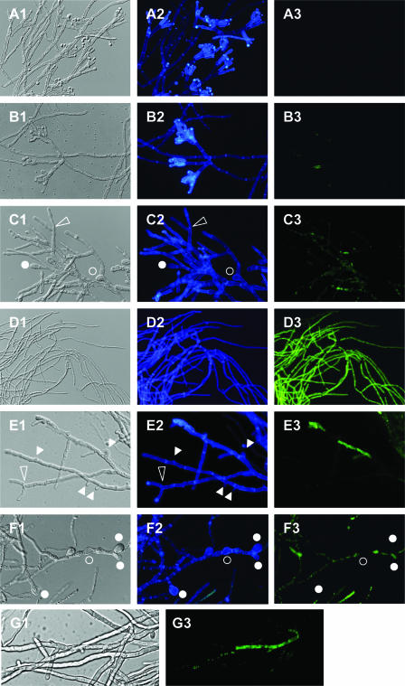
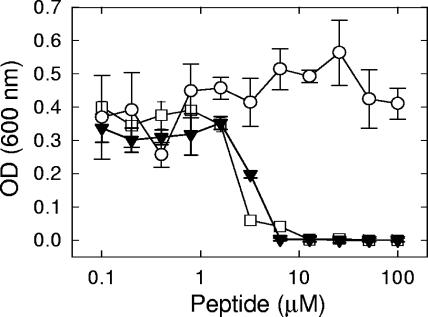
The small antimicrobial peptide PAF26 (Ac-RKKWFW-NH(2)) has been identified by a combinatorial approach and shows preferential activity toward filamentous fungi. In this work, we investigated the mode of action and inhibitory effects of PAF26 on the fungus Penicillium digitatum. The dye Sytox Green was used to demonstrate that PAF26 induced cell permeation. However, microscopic observations showed that sub-MIC concentrations of PAF26 produced both alterations of hyphal morphology (such as altered polar growth and branching) and chitin deposition in areas of no detectable permeation. Analysis of dose-response curves of inhibition and permeation suggested that growth inhibition is not solely a consequence of permeation. In order to shed light on the mode of PAF26 action, its antifungal properties were compared with those of melittin, a well-known pore-forming peptide that kills through cytolysis. While the 50% inhibitory concentrations and MICs of the two peptides against P. digitatum mycelium were comparable, they differed markedly in their fungicidal activities toward conidia and their hemolytic activities toward human red blood cells. Kinetic studies showed that melittin quickly induced Penicillium cell permeation, while PAF26-induced Sytox Green uptake was significantly slower and less efficient. Therefore, the ultimate growth inhibition and morphological alterations induced by PAF26 for P. digitatum are not likely a result of conventional pore formation. Fluorescently labeled PAF26 was used to demonstrate its specific in vivo interaction and translocation inside germ tubes and hyphal cells, at concentrations as low as 0.3 muM (20 times below the MIC), at which no inhibitory, morphological, or permeation effects were observed. Interestingly, internalized PAF26 could bind to cellular RNAs, since in vitro nonspecific RNA binding activity of PAF26 was demonstrated by electrophoretic mobility shift assays. We propose that PAF26 is a short, de novo-designed penetratin-type peptide that has multiple detrimental effects on target fungi, which ultimately result in permeation and killing.


Figures

FIG. 1.
FIG. 1.
Fluorescence microscopy of P. digitatum mycelium treated with PAF26. Mycelium was incubated in 5% PDB at 24°C without peptide (panels A) or at PAF26 final concentrations of 1 μM (B and E), 3 μM (C, F, and G), or 15 μM (D) for 48 h. After incubation, samples were stained with CFW and SG. Panels represent bright-field images (left; suffix 1 in panel lettering), blue fluorescence indicative of CFW staining (center; suffix 2), and green fluorescence indicative of SG uptake (right; suffix 3) for the same fields. Micrographs A to F were captured with a light fluorescence microscope, while micrograph G was captured with a confocal laser microscope. Circles indicate swollen cells either at the terminus of hyphae (filled circles) or within hyphae (open circles). Open triangles indicate dichotomous tip branching; filled triangles indicate lateral aborted branching.
FIG. 2.
FIG. 2.
Effects on growth and SG uptake by P. digitatum mycelium of peptides PAF26 (top), melittin (center), and mel.subK7I (bottom). Mycelium was incubated with peptides in 5% PDB at 24°C for 24 h. After incubation, SG was added and incubated for an additional 4 h. Dose-response curves show the increase in the OD at 492 nm (left axis; open circles) and the fluorescence intensity (FI) at 538 nm (right axis; filled triangles) after peptide addition for each peptide concentration (μM). Plotted values are means ± standard deviations of three replicate samples. a.u., arbitrary units.
FIG. 3.
FIG. 3.
Time course of SG uptake by P. digitatum mycelium treated with peptide PAF26 (top), melittin (center), or mel.subK7I (bottom). Mycelium was simultaneously exposed to peptides (at different concentrations) and to 0.2 μM SG in 5% PDB. Immediately after peptide and SG addition, fluorescence at 538 nm was recorded at 2-min intervals for 180 min. Curves show mean fluorescence intensity (FI) values in triplicate samples at 10-min intervals for 0.5 (filled circles), 1 (open circles), 3 (filled triangles), 6 (open triangles), 8 (filled squares), 10 (open squares), and 15 (filled diamonds) μM of the different peptides.
FIG. 4.
FIG. 4.
Killing kinetics of P. digitatum conidia by synthetic peptides. Conidia (104/ml) were incubated in water in the absence of peptide (filled circles) or in the presence of 30 μM PAF26 (open triangles) or 30 μM melittin (open squares). Aliquots were removed at different time points from 15 min to 16 h, diluted, and plated on PDA in the absence of peptide to assay for CFU recovery. Values are means ± standard deviations of three replicate samples.
FIG. 5.
FIG. 5.
Dose-response curves of the effects on the growth of P. digitatum of peptides PAF26 (filled triangles) and FITC-PAF26 (open squares) and the fluorophore FITC (open circles). Conidia (2.5 × 10⁴/ml) in 5% PDB were exposed to different concentrations of compounds and incubated at 24°C. Dose-response curves show mean ODs at 600 nm ± standard deviations after 48 h of incubation. The IC50s of PAF26 and FITC-PAF26 did not differ significantly (P > 0.05).
FIG. 6.
FIG. 6.
Interaction of FITC-PAF26 with P. digitatum. (A and B) Confocal laser microscope localization of FITC-PAF26 in P. digitatum conidia. Quiescent conidia were exposed in distilled water to 30 μM FITC-PAF26 for 30 min (A) or 16 h (B). Bright-field (panels 1), fluorescence (panels 2), and overlay (panels 3) micrographs are shown. Arrowhead points to a minimal fluorescence signal observed inside the spore. (C to F) Confocal laser microscope localization of FITC-PAF26 in germ tubes and hyphae of P. digitatum. Mycelium was exposed in distilled water to 0.3 μM FITC-PAF26 for less than 2 min (C and D), approximately 5 min (E), or approximately 15 min (F). (G) Confocal laser microscope image of P. digitatum mycelium simultaneously exposed to 0.3 μM FITC and 0.3 μM PAF26 for approximately 15 min. In panels C, bright-field, fluorescence, and overlay micrographs are shown as described above, whereas in panels D to G, only bright-field and fluorescence micrographs are shown.
FIG. 7.
FIG. 7.
Analysis of RNA-binding properties of PAF26 by EMSA. Yeast tRNA (250 ng) was incubated with no peptide (rightmost lane) or with increasing amounts of peptide (given above the gel) and analyzed by EMSA. The positions of the origin (O) and of free tRNA are indicated on the left.

Similar articles

Cited by

References

    1. Broekaert, W. F., F. R. G. Terras, B. P. A. Cammue, and J. Vanderleyden. 1990. An automated quantitative assay for fungal growth-inhibition. FEMS Microbiol. Lett. 69:55-59.
    1. Brogden, K. A. 2005. Antimicrobial peptides: pore formers or metabolic inhibitors in bacteria? Nat. Rev. Microbiol. 3:238-250. - PubMed
    1. Eckert, R., F. X. Qi, D. K. Yarbrough, J. He, M. H. Anderson, and W. Y. Shi. 2006. Adding selectivity to antimicrobial peptides: rational design of a multidomain peptide against Pseudomonas spp. Antimicrob. Agents Chemother. 50:1480-1488. - PMC - PubMed
    1. Epand, R. M., and H. J. Vogel. 1999. Diversity of antimicrobial peptides and their mechanisms of action. Biochim. Biophys. Acta 1462:11-28. - PubMed
    1. Gonzalez, C. F., E. M. Provin, L. Zhu, and D. J. Ebbole. 2002. Independent and synergistic activity of synthetic peptides against thiabendazole-resistant Fusarium sambucinum. Phytopathology 92:917-924. - PubMed

Publication types