. 2006 Nov;119(3):421-9.
doi: 10.1111/j.1365-2567.2006.02453.x.

Glucocorticoid-induced tumour necrosis factor receptor family related protein (GITR) mediates inflammatory activation of macrophages that can destabilize atherosclerotic plaques

Affiliations

Glucocorticoid-induced tumour necrosis factor receptor family related protein (GITR) mediates inflammatory activation of macrophages that can destabilize atherosclerotic plaques

Won-Jung Kim et al. Immunology. 2006 Nov.

Abstract

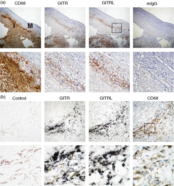
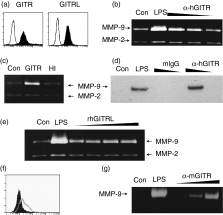
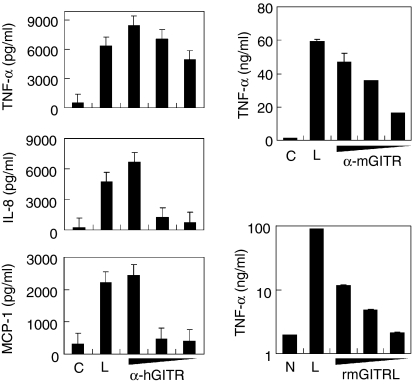
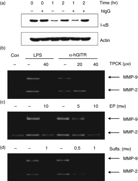
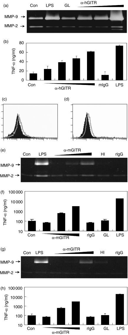
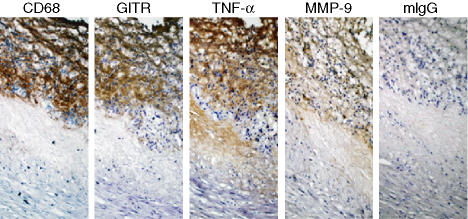
Glucocorticoid-induced tumour necrosis factor receptor family related protein (GITR) is the 18th member of the tumour necrosis factor receptor superfamily (TNFRSF18) and is known to interact with its cognate ligand GITRL (TNFSF18). We investigated the potential role of GITR in the pro-inflammatory activation of macrophages. Immunohistochemistry and in situ hybridization analyses of human atherosclerotic plaques demonstrated that GITR and its ligand are expressed mainly in lipid-rich macrophages. We then investigated the role of GITR in human and mouse monocyte/macrophage functions. Stimulation of GITR caused nuclear factor (NF)-kappaB-dependent activation of matrix metalloproteinase-9 (MMP-9) and pro-inflammatory cytokine expression in both the human and mouse monocytic/macrophage cell lines, THP-1 and RAW264.7, respectively. These cellular responses were also observed when the THP-1 cells were treated with phorbol-12 myristate-13 acetate (PMA), which is known to induce macrophage differentiation. To demonstrate that these responses are not restricted to cultured cell lines, we tested primary macrophages. Both peritoneal and bone marrow-derived macrophages responded to GITR stimulation with induction of MMP-9 and tumour necrosis factor-alpha (TNF-alpha). Furthermore, the GITR staining pattern overlapped with those of MMP-9 and TNF-alpha in atherosclerotic plaques. These data indicate that GITR-mediated macrophage activation may promote atherogenesis via the induction of pro-atherogenic cytokines/chemokines, and destabilize the atherosclerotic plaques via the induction of the matrix-degrading enzyme, MMP-9.


Figures

Figure 1
Figure 1
Macrophages express GITR and GITRL in the atherosclerotic plaques. (a) Plaque sections were stained with antibodies against CD68, GITR, or GITRL. Isotype-matching control antibody (mouse IgG1) was used to demonstrate the specificity of the staining. Low magnification (× 100) pictures of consecutive sections of atherosclerotic plaque are shown in the upper panel and high magnification (× 400) pictures are shown in the lower panel. I, intima; M, media. (b) Serial sections of an atherosclerotic plaque were used for in situ hybridization using GITR- or GITRL-specific probes or a non-specific control probe as described in the Materials and methods section. The hybridization was visualized as dark purple. For comparison, an adjacent section was immunostained using anti-CD68 mAb (brown colour). Magnifications are × 400 for the upper panel and × 1000 for the lower panel.
Figure 2
Figure 2
Stimulation of GITR induces MMP-9 expression in monocyte/macrophage cell lines. (a) THP-1 cells were stained with anti-GITR or anti-GITRL mAbs as indicated. Fluorescence profiles obtained from specific staining (filled area) and background staining (open area, stained with isotype-matching control antibody) are compared. (b) THP-1 cells were stimulated with anti-hGITR mAb immobilized at 20, 6 and 2 μg/ml concentrations. Culture supernatants were collected over a 24 hr period to measure MMP-9 activity using gelatin zymogram. (c) THP-1 cells were stimulated with immobilized anti-GITR mAb (10 μg/ml) that had been pretreated with (HI) or without (GITR) heat (95° for 30 min). (d) THP-1 cells were stimulated with anti-GITR mAb or mIgG immobilized at 20 and 2 μg/ml concentrations. Culture supernatants were collected after 24 hr and condensed approximately 10-fold before Western blot analysis with anti-MMP-9 mAb. (e) THP-1 cells were stimulated with 0·1, 0·3, 1 and 3 μg/ml of rhGITRL for 24 hr before gelatin zymogram. (f) RAW 264.7 cells were stained with anti-GITR mAb. Histograms from specific staining (open area) and background staining (filled area, stained with isotype-matching control antibody) are compared. (g) RAW264.7 cells were stimulated with anti-mGITR mAb which was added to culture medium at 1, 10 and 30 μg/ml concentrations. Culture supernatants were collected after 48 hr for gelatin zymogram. As a positive control, the cells were treated with 1 μg/ml lipopolysaccharide (LPS). Con, no treatment control.
Figure 3
Figure 3
Stimulation of GITR induces cytokine/chemokine expression in monocyte/macrophage cell lines. (a) THP-1 cells were stimulated with immobilized anti-hGITR mAb or mIgG. Culture supernatants were collected 24 hr after activation and cytokine concentrations were measured using ELISA. Concentrations used for the immobilization were 200, 20 and 2 μg/ml for the anti-GITR mAb and 200 μg/ml for mIgG. (b,c) RAW264.7 cells were stimulated with anti-mGITR mAb added to the culture medium at 30, 10 and 1 μg/ml concentrations or 30 μg/ml of rat IgG (b) or rmGITRL added at 10, 3 and 1 μg/ml concentrations (c). Culture supernatants were collected 48 hr after activation and TNF-α concentrations were measured using ELISA. C, treatment with isotype-matching control antibody; N, no treatment control; L, treatment with 1 μg/ml lipopolysaccharide as a positive control. Mean values of triplicate measurements ± SD are shown.
Figure 4
Figure 4
Stimulation of GITR induces the NF-κB activation, which is required for MMP-9 induction. (a) THP-1 cells were preincubated with or without human IgG (20 μg/ml) for 30 min to block FcRs. These cells were then stimulated with immobilized anti-GITR mAb (20 μg/ml). Whole cell lysates were then collected 0, 1 or 2 hr after the activation and IκB levels were measured by Western blot analysis. Actin levels were measured as a protein loading control. (b–d), THP-1 cells were activated with immobilized anti-GITR mAb in the presence or absence of NF-κB inhibitors TPCK (b), ethyl pyruvate (c), and sulphasalazine (d). Culture supernatants were collected after 24 hr for the measurement of MMP-9 activity using gelatin zymogram. Con, no treatment control; LPS, treatment with 1 μg/ml lipopolysaccharide as a positive control.
Figure 5
Figure 5
GITR stimulation activates macrophages to express MMP-9 and TNF-α. THP-1 cells were treated with PMA to differentiate into macrophages, as described in the Materials and methods section. Cells were then stimulated with 3 μg/ml rhGITRL (GL) or 3, 10 and 30 μg/ml anti-hGITR mAb, which was added to the culture medium. As a positive control, cells were stimulated with 1 μg/ml LPS. Culture supernatants were collected after 24 hr and MMP-9 levels were measured using gelatin zymogram (a) and TNF-α levels were measured using ELISA (b). Peritoneal macrophages (c, e, f) and bone marrow-derived macrophages (d, g, h) were analysed. Fluorescence profiles of GITR specific staining (open area) were compared with that obtained using isotype-matching control antibody (filled area) (c, d). Cells were stimulated with anti-mGITR mAb added to the culture medium at 3, 10 and 30 μg/ml concentrations or 3 μg/ml of rmGITRL (GL). As negative controls, cells were also stimulated with heat-inactivated (HI) anti-GITR mAb or rat IgG (rIgG) at 30 μg/ml. Culture supernatants were collected after 48 hr and MMP-9 levels were measured using gelatin zymogram (e, g). TNF-α levels were measured using ELISA (f, h).
Figure 6
Figure 6
Expression of TNF-α and MMP-9 co-localized with that of GITR in human atherosclerotic plaques. Consecutive sections of a human carotid artherosclerotic plaque were stained for the expression of CD68, GITR, TNF-α and MMP-9. Pictures were taken at × 400 magnification. As a negative control, mouse IgG1 was used for staining.

References

    1. Rus HG, Niculescu F, Vlaicu R. Tumor necrosis factor-alpha in human arterial wall with atherosclerosis. Atherosclerosis. 1991;89:247–54. - PubMed
    1. Kaartinen M, Penttila A, Kovanen PT. Mast cells in rupture-prone areas of human coronary atheromas produce and store TNF-α. Circulation. 1996;94:2787–92. - PubMed
    1. Mach F, Schonbeck U, Sukhova GK, Bourcier T, Bonnefoy JY, Pober JS, Libby P. Functional CD40 ligand is expressed on human vascular endothelial cells, smooth muscle cells, and macrophages: implications for CD40–CD40 ligand signaling in atherosclerosis. Proc Natl Acad Sci USA. 1997;94:1931–6. - PMC - PubMed
    1. Lee WH, Kim SH, Lee Y, Lee BB, Kwon B, Song H, Kwon BS, Park JE. Tumor necrosis factor receptor superfamily 14 is involved in atherogenesis by inducing proinflammatory cytokines and matrix metalloproteinases. Arterioscler Thromb Vasc Biol. 2001;21:2004–10. - PubMed
    1. Kim WJ, Lee WH. LIGHT is expressed in foam cells and involved in destabilization of atherosclerotic plaques through induction of matrix metalloproteinase-9 and IL-8. Immune Network. 2004;4:116–22.

Publication types

MeSH terms