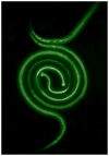
Strongyloides stercoralis: Amphidial neuron pair ASJ triggers significant resumption of development by infective larvae under host-mimicking in vitro conditions
- PMID: 17067579
- PMCID: PMC3091007
- DOI: 10.1016/j.exppara.2006.08.010
Strongyloides stercoralis: Amphidial neuron pair ASJ triggers significant resumption of development by infective larvae under host-mimicking in vitro conditions
Abstract
Resumption of development by infective larvae (L3i) of parasitic nematodes upon entering a host is a critical first step in establishing a parasitic relationship with a definitive host. It is also considered equivalent to exit from the dauer stage by the free-living nematode Caenorhabditis elegans. Initiation of feeding, an early event in this process, is induced in vitro in L3i of Strongyloides stercoralis, a parasite of humans, other primates and dogs, by culturing the larvae in DMEM with 10% canine serum and 5mM glutathione at 37 degrees C with 5% CO(2). Based on the developmental neurobiology of C. elegans, resumption of development by S. stercoralis L3i should be mediated, in part at least, by neurons homologous to the ASJ pair of C. elegans. To test this hypothesis, the ASJ neurons in S. stercoralis first-stage larvae (L1) were ablated with a laser microbeam. This resulted in a statistically significant (33%) reduction in the number of L3i that resumed feeding in culture. In a second expanded investigation, the thermosensitive ALD neurons, along with the ASJ neurons, were ablated, but there was no further decrease in the initiation of feeding by these worms compared to those in which only the ASJ pair was ablated.
Figures


References
-
- Ashton FT, Bhopale VM, Fine AE, Schad GA. Sensory neuro-anatomy of a skin-penetrating nematode parasite: Strongyloides stercoralis. I. Amphidial neurons. Journal of Comparative Neurology. 1995;357:281–295. - PubMed
-
- Ashton FT, Bhopale VM, Holt D, Smith G, Schad GA. Developmental switching in the parasitic nematode Strongyloides stercoralis is controlled by the ASF and ASI amphidial neurons. Journal of Parasitology. 1998;84:691–695. - PubMed
-
- Ashton FT, Li J, Schad GA. Chemo- and thermosensory neurons: structure and function in animal parasitic nematodes. Veterinary Parasitology. 1999;84:297–316. - PubMed
-
- Bargmann CI, Avery L. Laser killing of cells in Caenorhabditis elegans. In: Epstein HF, Shakes DC, editors. Caenorhabditis elegans: Modern Biological Analysis of an Organism. Academic Press; San Diego, CA: 1995. pp. 225–250.
-
- Bargmann CI, Horvitz HR. Control of larval development by chemosensory neurons in Caenorhabditis elegans. Science. 1991;251:1243–1246. - PubMed
Publication types
MeSH terms
Grants and funding
LinkOut - more resources
Full Text Sources

