Comparative Study
. 2006;30(5):403-11.
doi: 10.1016/j.cdp.2006.07.008. Epub 2006 Oct 25.

Cyclic adenosine monophosphate-dependent cell type-specific modulation of mitogenic signaling by retinoids in normal and neoplastic lung cells

Affiliations
Comparative Study

Cyclic adenosine monophosphate-dependent cell type-specific modulation of mitogenic signaling by retinoids in normal and neoplastic lung cells

Hussein A N Al-Wadei et al. Cancer Detect Prev. 2006.

Abstract

Background: Lung cancer is the leading cause of cancer death worldwide. A diet rich in fruit and vegetables has been shown to reduce the lung cancer risk. However, clinical trials with beta-carotene and retinoids have disappointed, resulted in increased mortality from lung cancer and cardiovascular disease.

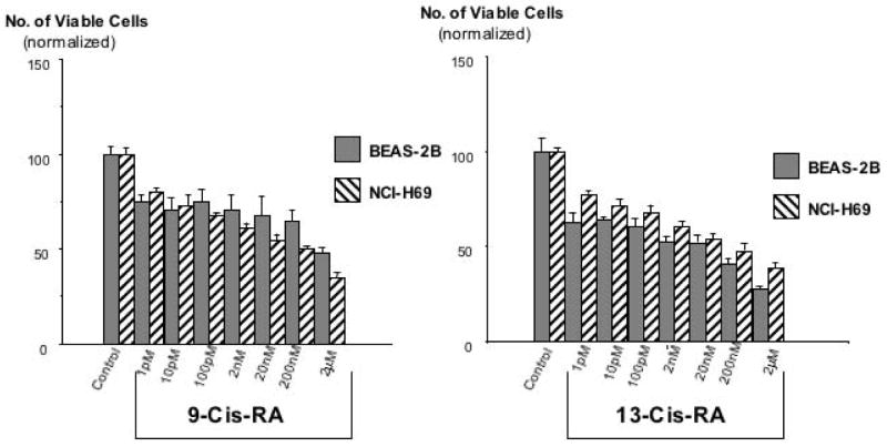
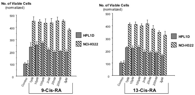
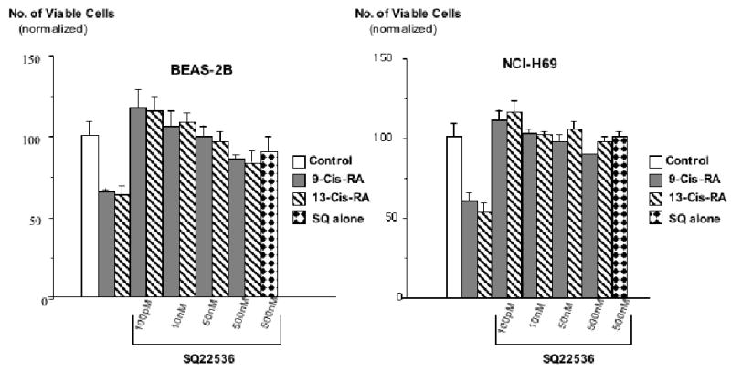
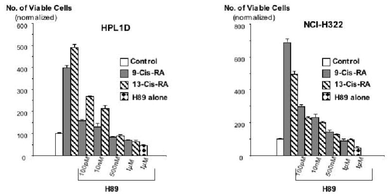
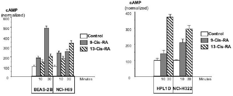
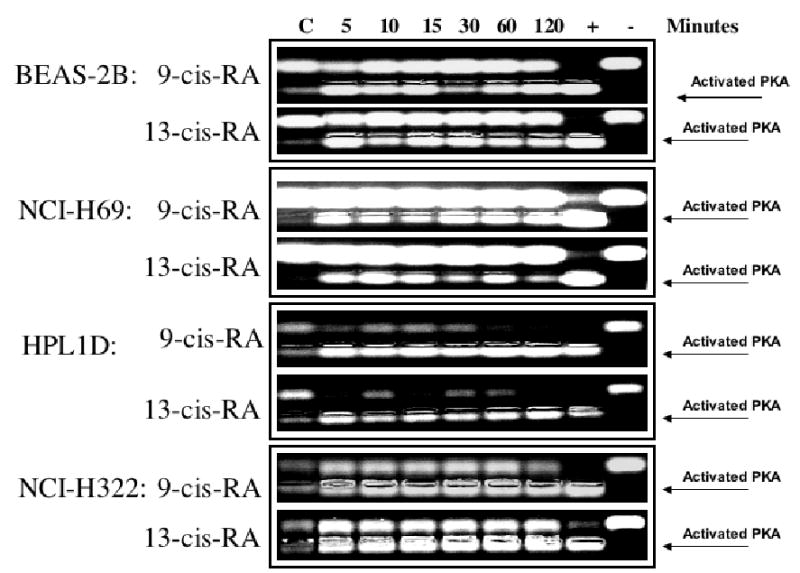
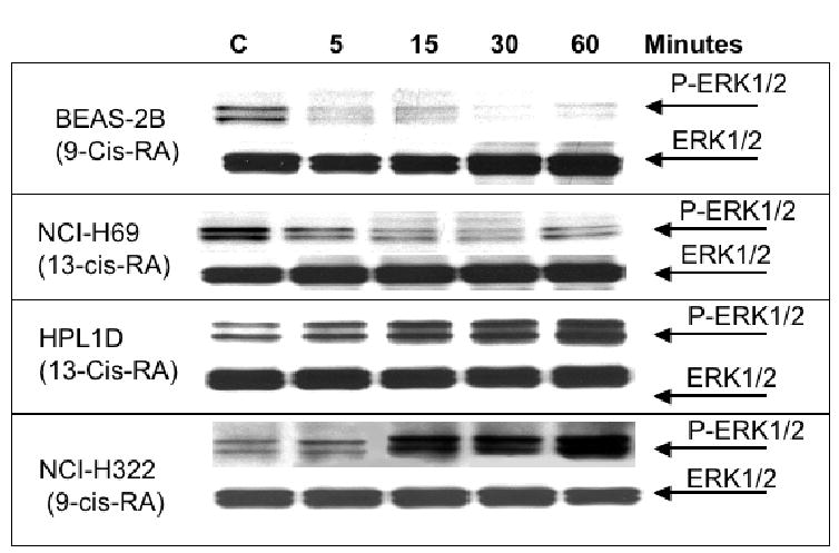
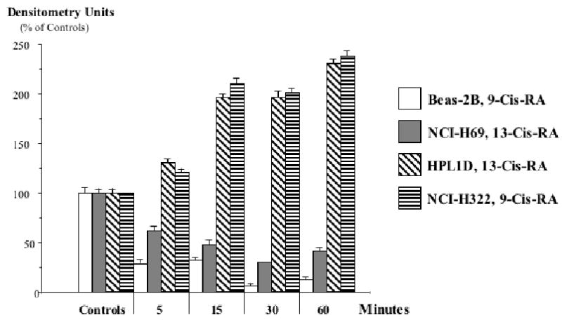
Methods: We have investigated the effects of the two major retinol metabolites, 9-cis-retinoic acid (9-Cis-RA), and 13-cis-retinoic acid (13-Cis-RA), on cell proliferation (MTT assays), intracellular cAMP (cAMP immunoassays), PKA activation (non-radioactive PKA activation assays), and ERK1/2 phosphorylation (Western blots) in immortalized human small airway epithelial cells, HPL1D, a human lung adenocarcinoma cell line, NCI-H322, immortalized human bronchial epithelial cells, BEAS-2B, and in the human small cell lung carcinoma cell line, NCI-H69.

Results: Both retinoids increased intracellular cAMP and PKA activation in all cell lines. In BEAS-2B and NCI-H69 cells, the stimulation of cAMP/PKA reduced the phosphorylation of ERK1/2 and inhibited cell proliferation whereas phosphorylation of ERK1/2 and cell proliferation were increased in HPL1D and NCI-H322 cells.

Conclusions: Our data have identified a novel mechanism of action of 9-Cis-RA and 13-Cis-RA: activation of PKA in response to increased cAMP. The observed stimulation of cAMP/PKA may inhibit the development of small cell lung carcinoma and other tumors derived from large airway epithelia whereas it may selectively promote the development of lung tumors derived from small airway epithelial cells, such as adenocarcinoma.


Figures

Figure 1
Figure 1
Results of MTT assays for the assessment of cell proliferation in the human large airway epithelial cells, BEAS-2B, and the human small cell lung carcinoma cells, NCI-H69, after exposure for 72 hours to 9-Cis-RA or 13-Cis-RA. Both cell lines responded with a concentration-dependent reduction in cell numbers. The observed responses were highly significant (P<0.001) at all retinoid concentrations tested. Data are mean values and standard errors of four samples per treatment group. Each assay was repeated twice and yielded similar data.
Figure 2
Figure 2
Results of MTT assay for the assessment of cell proliferation in the human small airway epithelial cells, HPL1D, and the human lung adenocarcinoma cells, NCI-H322 after exposure for 48 hours to 9-Cis-RA or 13-Cis-RA. Both cell lines responded with an increase in cell numbers. The observed responses were highly significant (P<0.001) at all retinoid concentrations tested. Data are mean values and standard errors of four samples per treatment group. Each assay was repeated twice with similar results.
Figure 3
Figure 3
Results of MTT assays, illustrating the effects of pre-incubation (10 minutes) with the adenylate cyclase inhibitor, SQ22536, on the response of large airway epithelial cells BEAS-2B and small cell lung carcinoma cells NCI-H69 cells to 9-Cis-RA or 13-Cis-RA. The retinoid-induced reduction of cell proliferation was completely abrogated by all concentrations of SQ22536 in both cell lines. These effects of SQ22536 were highly significant (P<0.001). Data are mean values and standard errors of four samples per treatment group. Each assay was repeated twice and yielded similar results.
Figure 4
Figure 4
Results of MTT assays, illustrating the effects of pre-incubation (20 minutes) with the PKA inhibitor, H89, on the responses of small airway epithelial cells HPL1D and lung adenocarcinoma cells NCI-H322 to 9-Cis-RA or 13-Cis-RA. The retinoid-induced stimulation of cell proliferation was inhibited by H89 in a concentration-dependent manner. These effects of H89 were highly significant (P<0.001). Data are mean values and standard errors of four samples per treatment group. Each assay was repeated twice and yielded similar results.
Figure 5
Figure 5
Effects of 9-Cis-RA or 13-Cis-RA on intracellular cAMP as assessed by a cAMP immunoassay in large airway epithelial cells BEAS-2B, small cell lung carcinoma cells NCI-H69, small airway epithelial cells HPL1D, or lung adenocarcinoma cells NCI-H322 after 10 and 30 minutes of incubation. Each retinoid significantly (P<0.001) increased the concentration of cAMP in each cell line. Data are mean values and standard errors of triplicate samples per treatment group. Each assay was repeated twice and yielded similar results.
Figure 6
Figure 6
PKA activation over time in response to 9-Cis-RA or 13-Cis-RA in large airway epithelial cells BEAS-2B, small cell lung carcinoma cells NCI-H69, small airway epithelial cells HPL1D, and lung adenocarcinoma cells NCI-H322 as assessed by a non-radioactive PKA activation assay. Each retinoid activated PKA in each cell line at all time intervals tested.
Figure 7
Figure 7
Western blot, indicating the effects of 9-Cis-RA or 13-Cis-RA on the expression of phosphorylated and unphosphorylated ERK1/2 proteins. Each retinoid decreased the expression of P-ERKL1/2 in large airway epithelial cells BEAS-2B and small cell lung carcinoma cells NCI-H69. By contrast, small airway epithelial cells HPL1D and lung adenocarcinoma cells NCI-H322 responded with an increase in P-ERK1/2 expression.
Figure 8
Figure 8
Quantitative assessment by densitometry of P-ERK1/2 expression in the Western blots of Figure 7. The observed decrease in P-ERK1/2 caused by 9-Cis-RA or 13-Cis-RA in large airway epithelial cells BEAS-2B and small cell lung carcinoma cells NCI-H69 was highly significant (p<0.001) at all time intervals tested, as was the increase in P-ERK1/2 expression observed in small airway epithelial cells HPL1D and lung adenocarcinoma cells NCI-H322. Data are mean values and standard errors of five densitometric readings per band.

References

    1. Weir HK, Thun MJ, Hankey BF, Ries LA, Howe HL, Wingo PA, et al. Annual report to the nation on the status of cancer, 1975–2000, featuring the uses of surveillance data for cancer prevention and control. J Natl Cancer Inst. 2003;95:1276–1299. - PubMed
    1. Ezzati M, Henley SJ, Lopez AD, Thun MJ. Role of smoking in global and regional cancer epidemiology: current patterns and data needs. Int J Cancer. 2005;116:963–971. - PubMed
    1. Garber ME, Troyanskaya OG, Schluens K, Petersen S, Thaesler Z, Pacyna-Gengelbach M, et al. Diversity of gene expression in adenocarcinoma of the lung. Proc Natl Acad Sci U S A. 2001;98:13784–13789. - PMC - PubMed
    1. Smith-Warner SA, Spiegelman D, Yaun SS, Albanes D, Beeson WL, van den Brandt PA, et al. Fruits, vegetables and lung cancer: a pooled analysis of cohort studies. Int J Cancer. 2003;107:1001–1011. - PubMed
    1. Miller AB, Altenburg HP, Bueno-de-Mesquita B, Boshuizen HC, Agudo A, Berrino F, et al. Fruits and vegetables and lung cancer: findings from the European prospective investigation into cancer and nutrition. Int J Cancer. 2004;108:269–276. - PubMed

Publication types

MeSH terms