Review
. 2006;34(5):648-55.
doi: 10.1080/01926230600865523.

Enhanced histopathology of the spleen

Affiliations
Review

Enhanced histopathology of the spleen

Susan A Elmore. Toxicol Pathol. 2006.

Abstract

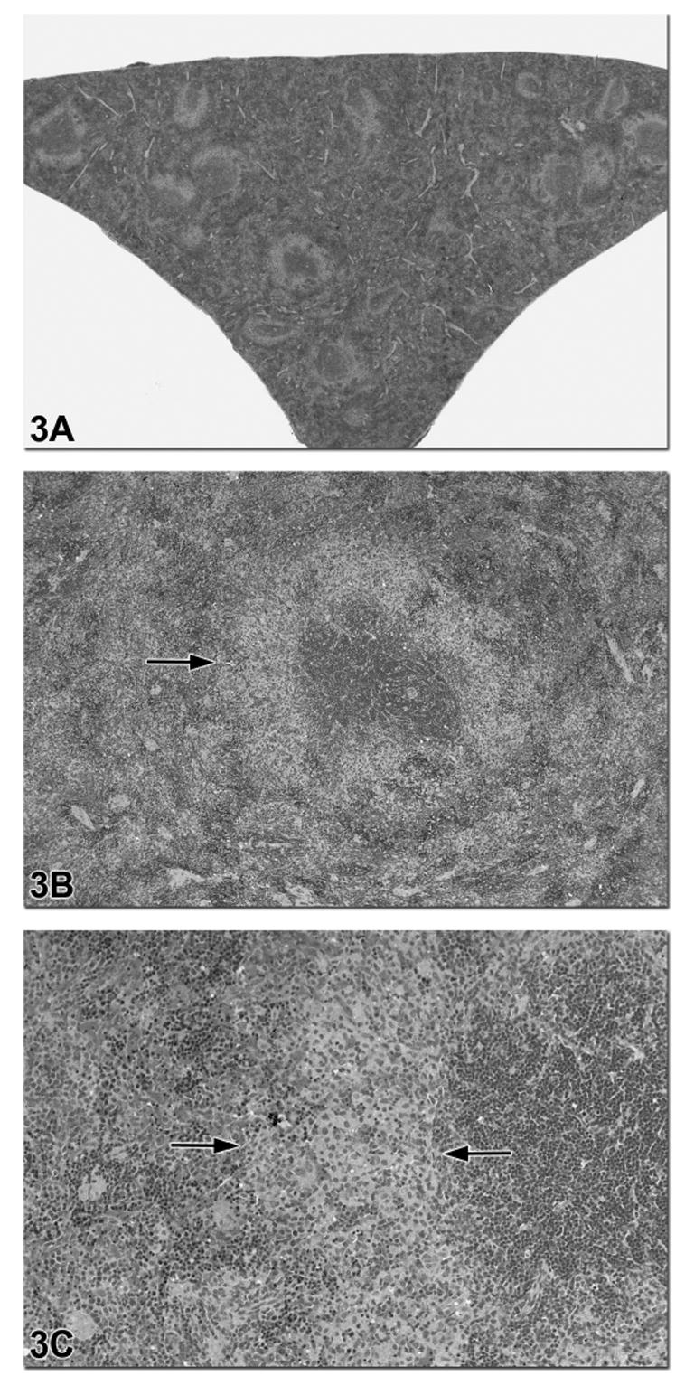
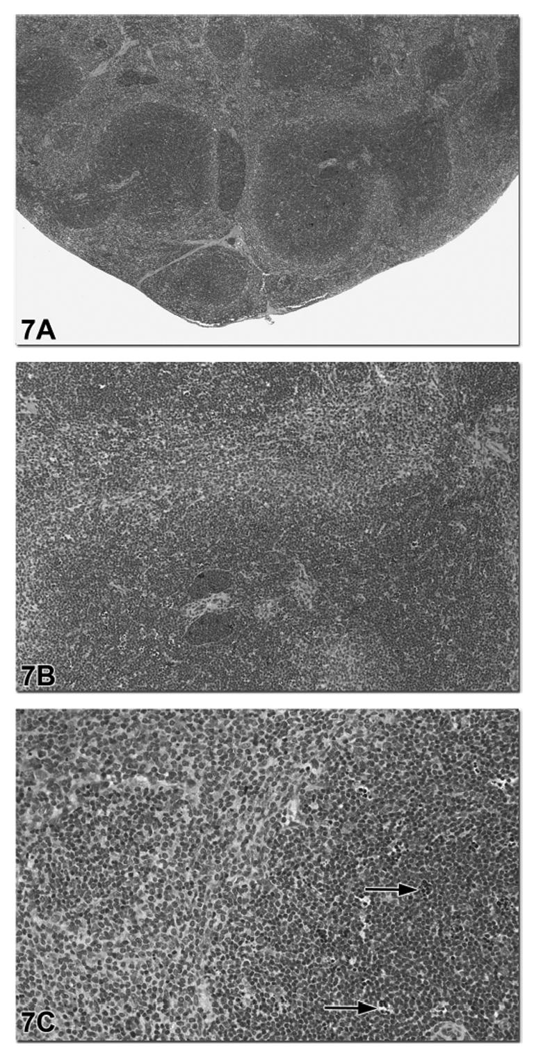
The spleen is the largest secondary lymphoid organ, is considered the draining site for compounds that are administered intravenously, and is therefore considered an important organ to evaluate for treatment-related lesions. Due to the presence of B and T lymphocytes, the immunotoxic effects of xenobiotics or their metabolites on these cell populations may be reflected in the spleen. Therefore it is one of the recommended organs to evaluate for enhanced histopathology of the immune system. The two major functional zones of the spleen are the hematogenous red pulp and the lymphoid white pulp (periarteriolar sheaths, follicles and marginal zones). For enhanced histopathology, these splenic compartments should be evaluated separately for changes in size and cellularity, and descriptive rather than interpretive terminology should be used to characterize any changes (Haley et al., 2005). Moreover, germinal center development within the lymphoid follicles should be noted as increased or decreased.


Figures

Figure 1
Figure 1
Normal spleen from a control rat is depicted in Figures 1a–c. In Figure 1a the arrowhead indicates the PALS region of the spleen surrounding the central artery. This is the darkest region of the white pulp due to the presence of predominately small lymphocytes. The short arrow indicates a lymphoid follicle whereas the long arrow indicates the marginal zone that surrounds both the PALS and the follicle. In this control spleen, apoptotic cells are rare within these three regions of the white pulp. The arrows in Figures 1b and 1c indicate two small heterochromatic cell fragments, indicative of apoptotic bodies.
Figure 2
Figure 2
Splenic tissue from a 30-day-old Sprague Dawley rat treated with dexamethasone is depicted in Figures 2a–d. At low magnification (Figure 2a) the PALS region appears “moth-eaten” due to a moderate degree of scattered lymphocyte apoptosis (arrow). The arrowhead in Figure 2a indicates the presence of apoptotic cells within the periphery of the marginal zone. Figures 2b and 2c show higher magnifications of this PALS region. The arrows indicate tingible body macrophages with cytoplasmic engulfed apoptotic debris and the arrowhead indicates free apoptotic bodies. Figure 2d is a higher magnification of the region depicted in Figure 2a with an arrowhead. This image illustrates a region of the peripheral marginal zone with apoptotic bodies and tingible body macrophages.
Figure 3
Figure 3
Figures 3a–c are images of a spleen from a 90-day-old F344 rat treated with riddelline. There is diffuse and severe decreased cellularity of the B lymphocytes in the marginal zone (arrows in 3b and 3c). Higher magnification in Figure 3b shows that the T cell-rich PALS regions and B cell-rich follicles are not affected. The area of B lymphocyte depletion in the marginal zone is paler staining due to the presence of macrophages (Figure 3c).
Figure 4
Figure 4
Figures 4a and 4b are low and high magnifications images of a spleen from a male F344 rat in a N,N-dimethyl-p-toluidine subchronic study. Figures 4c and 4d are low and high magnification images from an age- and sex-matched control rat with prominent marginal zones. The spleen from the treated rat had a diagnosis of decreased white pulp area and cellularity and decreased red pulp area and cellularity. Figure 4b illustrates that all compartments of the white pulp (PALS, follicles, marginal zone) were similarly affected.
Figure 5
Figure 5
This spleen is from a female B6C3F1 mouse that was treated with 800 mg/kg bodyweight of the antiviral drug azidothymidine (AZT) plus methadone HCL for 90 days. This treatment resulted in decreased cellularity of both the white pulp and red pulp. The capsular surface has a distinctive scalloped appearance, in this case indicative of a decrease in organ size due to the marked loss of lymphocytes and red blood cells. The higher magnification in 5b illustrates the variable decrease in cellularity in the PALS, marginal zones and follicles. As with all enhanced histopathology evaluations, each compartment should be evaluated separately and compared with controls.
Figure 6
Figure 6
The low and high magnification images of the spleen in Figures 6a and 6b are from a 90-day-old male B6C3F1 mouse treated with citral and an age-and sex-matched control from the same study is provided in Figures 6c and 6d for comparison. In this study, the diagnosis for the spleen in Figure 6a was a mild increase in lymphocyte cellularity. At high magnification (Figure 6b) the PALS regions and marginal zones have increased cellularity and there is a relative decrease in the area of red pulp due to the expansion of the white pulp as well as a decrease in hematopoietic precursor cells. This type of lesion would have to be distinguished from neoplasia. This example illustrates the need to compare a lesion with appropriate controls in order to determine the range of normal histology and to devise an appropriate grading scheme.
Figure 7
Figure 7
This spleen is from a male B6C3F1 mouse in a subchronic androstenedione study. The diagnosis was a mild increase in lymphocyte cellularity and mild lymphocyte apoptosis based on an appropriate grading scheme for this study. Figure 7a shows the expansion of the white pulp with a relative decrease in the area of red pulp. Figure 7b shows that the increase in lymphocyte cellularity involves the T cell-rich PALS regions. Within these regions of lymphocyte hypercellularity there was scattered lymphocyte apoptosis with tingible body macrophages (arrows, Figure 7c).

References

    1. Cesta MF. Normal structure, function, and histology of the spleen. Toxicol Pathol. 2006;34:455–465. - PubMed
    1. Germolec DR, Kashon M, Nyska A, Kuper CF, Portier C, Kommineni C, Johnson KA, Luster MI. The accuracy of extended histopathology to detect immunotoxic chemicals. Toxicol Sci. 2004;82:504–14. - PubMed
    1. Gopinath C. Pathology of toxic effects on the immune system. Inflamm Res. 1996;45(Suppl 2):S74–S8. - PubMed
    1. Haley P, Perry R, Ennulat D, Frame S, Johnson C, Lapointe JM, Nyska A, Snyder P, Walker D, Walter G. STP position paper: best practice guideline for the routine pathology evaluation of the immune system. Toxicol Pathol. 2005;33:404–7. - PubMed
    1. Harleman JH. Approaches to the identification and recording of findings in the lymphoreticular organs indicative for immunotoxicity in regulatory type toxicity studies. Toxicology. 2000;142:213–9. - PubMed

Publication types