. 2007 Feb;292(2):G590-8.
doi: 10.1152/ajpgi.00252.2006. Epub 2006 Oct 26.

Mechanism of glucocorticoid regulation of the intestinal tight junction barrier

Affiliations

Mechanism of glucocorticoid regulation of the intestinal tight junction barrier

Michel A Boivin et al. Am J Physiol Gastrointest Liver Physiol. 2007 Feb.

Abstract

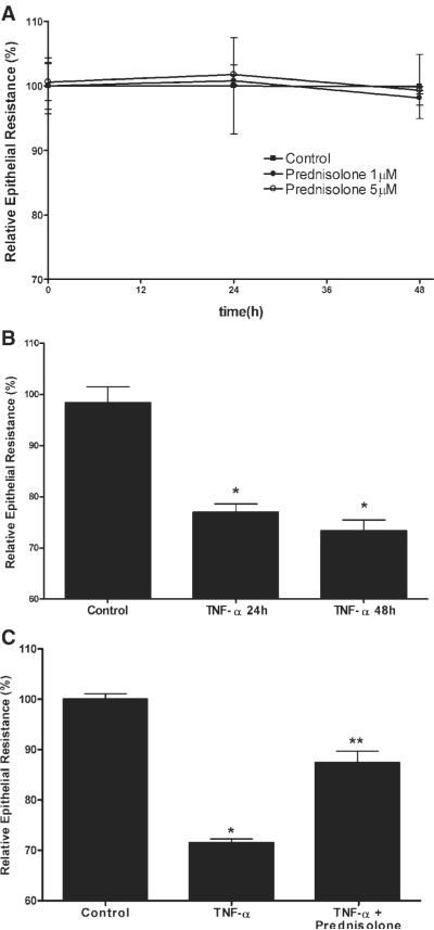
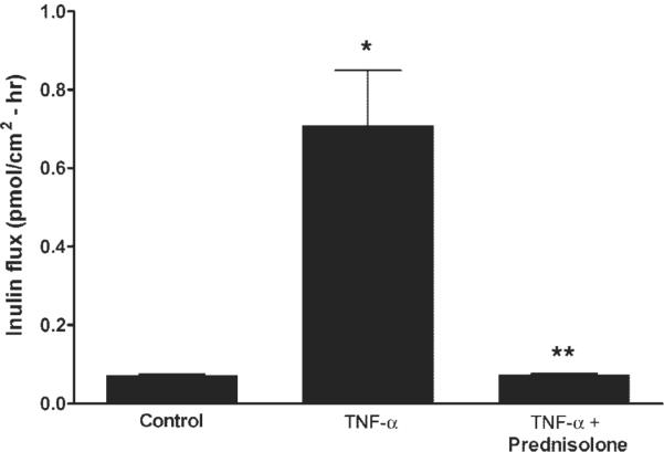
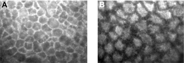
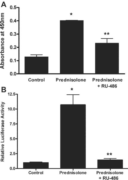
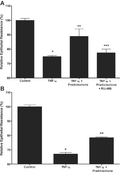
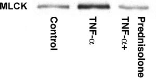
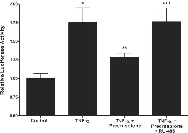
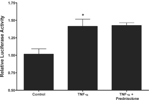
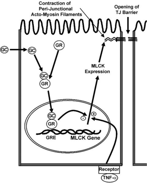
A defective intestinal epithelial tight junction (TJ) barrier has been proposed as an important pathogenic factor contributing to the intestinal inflammation of Crohn's disease. Glucocorticoids are first-line therapeutic agents for the treatment of moderate to severe Crohn's disease. Glucocorticoid treatment has been shown to induce retightening of the intestinal TJ barrier defect in Crohn's disease patients. However, the mechanisms that mediate the glucocorticoid therapeutic action on intestinal TJ barrier function remain unknown. The aim of this study was to elucidate the mechanism of glucocorticoid modulation of the intestinal epithelial TJ barrier using an in vitro model system. Filter-grown Caco-2 intestinal epithelial cells were used as an in vitro model to examine the effects of glucocorticoids on basal intestinal epithelial TJ barrier function and on TNF-alpha-induced disruption of the TJ barrier. Glucocorticoids (prednisolone and dexamethasone) did not have a significant effect on baseline Caco-2 TJ barrier function but prevented the TNF-alpha-induced increase in Caco-2 TJ permeability. The glucocorticoid protective effect against the TNF-alpha-induced increase in Caco-2 TJ permeability required activation of the glucocorticoid receptor (GR) complex. The activation of the GR complex resulted in GR complex binding to the glucocorticoid response element (GRE) site on DNA and activation of a GR-responsive promoter. Glucocorticoids inhibited the TNF-alpha-induced increase in myosin light chain kinase (MLCK) protein expression, a key process mediating the TNF-alpha increase in intestinal TJ permeability. The glucocorticoid inhibition of the TNF-alpha-induced increase in MLCK protein expression was due to the binding of the GR complex to a GRE binding site on the MLCK promoter region suppressing the TNF-alpha-induced activation. Glucocorticoids inhibit the TNF-alpha-induced increase in Caco-2 TJ permeability. The prednisolone protective action was mediated by binding of activated GR complex to the GRE site on the MLCK promoter, suppressing the TNF-alpha-induced increase in MLCK gene activity, protein expression, and subsequent opening of the intestinal TJ barrier.


Figures

Fig. 1
Fig. 1
Effect of prednisolone on the baseline and TNF-α-induced decrease in Caco-2 transepithelial electrical resistance (TER). A: time course of prednisolone effect on baseline Caco-2 TER. Filter-grown intestinal epithelial cells were treated with prednisolone (●,1 μM; ◯,5 μM) or vehicle (∎) over a 48-h experimental period. B: time course of TNF-α effect on TER. Filter-grown Caco-2 monolayers were treated with TNF-α or vehicle over a 24- and 48-h period (*P < 0.01). C: effect of prednisolone on the TNF-α-induced decrease in Caco-2 TER. Epithelial monolayers were treated with vehicle (control), TNF-α (10 ng/ml), or TNF-α+ prednisolone (5 μM) for 48 h. *P < 0.01 compared with control; **P < 0.01 compared with TNF-α.
Fig. 2
Fig. 2
Effect of TNF-α and prednisolone on Caco-2 paracellular permeability. The paracellular marker [14C]inulin was added to the apical chamber of filter-grown Caco-2 cells. After 48 h, the basolateral compartment was sampled over a 1-h interval. Inulin permeability was determined in groups treated with vehicle (control), TNF-α (10 ng/ml), or TNF-α+ prednisolone (5 μM). *P < 0.01 compared with control; **P < 0.01 compared with TNF-α.
Fig. 3
Fig. 3
Prednisolone effect on glucocorticoid (GC) receptor (GR) localization. Filter-grown Caco-2 monolayers were immunolabeled as described in MATERIALS AND METHODS for determination of cytoplasmic to nuclear translocation of GR. A: control monolayers. B: Caco-2 cells after incubation with prednisolone (5 μM) for 30 min (magnification, ×400).
Fig. 4
Fig. 4
Prednisolone effect on GR binding to DNA and promoter activation. A: effect of prednisolone on GR binding to a consensus GC response element (GRE) DNA binding site using an ELISA-based GR DNA binding assay as described in MATERIALS AND METHODS. Cells were treated with vehicle (control) or prednisolone (5 μM for 30 min) or pretreated with RU-486 (0.5 μM for 30 min) followed by prednisolone treatment. *P < 0.01 compared with control; **P < 0.05 compared with prednisolone. B: effect of prednisolone on GRE-responsive secreted embryonic alkaline phosphatase (SEAP)-promoter activity. Caco-2 intestinal epithelial cells (transfected with pGRE-SEAP) were treated with vehicle (control), prednisolone (5 μM), or prednisolone + RU-486 (1 μM) for 24 h. *P < 0.05 compared with control; **P < 0.05 compared with prednisolone.
Fig. 5
Fig. 5
A: effect of RU-486 on prednisolone protection of the TNF-α-induced drop in Caco-2 TER. Filter-grown epithelial monolayers were treated with vehicle (control), TNF-α (10 ng/ml), TNF-α + prednisolone (5 μM), or TNF-α, prednisolone, and RU-486 (1 μM) over a 48-h period. B: effect of short exposure (6 h) time of TNF-α and prednisolone on Caco-2 TER. Caco-2 cells were incubated with vehicle, TNF-α 10 ng/ml, or TNF-α+ prednisolone (5 μM) for 6 h and then replaced with regular medium. The effect of 6-h exposure on Caco-2 TER was determined at 24 h. *P < 0.01 compared with control; **P < 0.01 compared with TNF; ***P < 0.01 compared with prednisolone.
Fig. 6
Fig. 6
Effect of TNF-α and prednisolone on MLCK protein expression. Caco-2 myosin light chain kinase (MLCK) expression was assessed by Western blotting; bands are localized at ~210 kDa. Experimental groups were treated with vehicle (control), TNF-α (10 ng/ml), or TNF-α+ prednisolone (5 μM) for 48 h.
Fig. 7
Fig. 7
Effect of prednisolone on the TNF-α-induced upregulation of MLCK promoter activity. The pGL3 vector (containing the full-length MLCK promoter with a luciferase reporter) was transfected into Caco-2 cells as described in MATERIALS AND METHODS. Cells were treated with vehicle (control), TNF-α (10 ng/ml), TNF-α+ prednisolone (5 μM), or TNF-α, prednisolone, and RU-486 (1 μM)for6h.*P < 0.01 compared with control; **P < 0.05 compared with TNF-α; ***P < 0.05 compared with TNF-α+ prednisolone.
Fig. 8
Fig. 8
Effect of site-directed mutation of the GRE region on TNF-α and prednisolone modulation of the MLCK promoter. The mutant MLCK promoter containing the mutation of the GRE region was generated and then transfected into filter-grown Caco-2 cells as described in MATERIALS AND METHODS. Transfected Caco-2 cells were treated with vehicle (control), TNF-α (10 ng/ml), or TNF-α+ prednisolone (5 μM)for6h.*P < 0.01 compared with control. No significant difference between TNF-α and TNF-α+ prednisolone groups.
Fig. 9
Fig. 9
Proposed mechanism of GC modulation of the TNF-α-induced increase in intestinal epithelial tight junction (TJ) permeability.

References

    1. Anderson JM, Van Itallie CM. Tight junctions and the molecular basis for regulation of paracellular permeability. Am J Physiol Gastrointest Liver Physiol. 1995;269:G467–G475. - PubMed
    1. Arnott ID, Kingstone K, Ghosh S. Abnormal intestinal permeability predicts relapse in inactive Crohn disease. Scand J Gastroenterol. 2000;35:1163–1169. - PubMed
    1. Blair SA, Kane SV, Clayburgh DR, Turner JR. Epithelial myosin light chain kinase expression and activity are upregulated in inflammatory bowel disease. Lab Invest. 2006;86:191–201. - PubMed
    1. Clayburgh DR, Barrett TA, Tang Y, Meddings JB, Van Eldik LJ, Watterson DM, Clarke LL, Mrsny RJ, Turner JR. Epithelial myosin light chain kinase-dependent barrier dysfunction mediates T cell activation-induced diarrhea in vivo. J Clin Invest. 2005;115:2702–2715. - PMC - PubMed
    1. Fanning AS, Ma TY, Anderson JM. Isolation and functional characterization of the actin binding region in the tight junction protein ZO-1. FASEB J. 2002;16:1835–1837. - PubMed

Publication types

MeSH terms