. 2006 Nov 7;103(45):16953-8.
doi: 10.1073/pnas.0601562103. Epub 2006 Oct 26.

Legionella pneumophila glucosyltransferase inhibits host elongation factor 1A

Affiliations

Legionella pneumophila glucosyltransferase inhibits host elongation factor 1A

Yury Belyi et al. Proc Natl Acad Sci U S A. .

Abstract

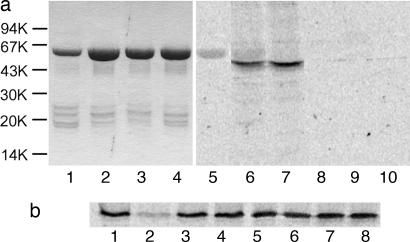
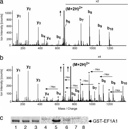
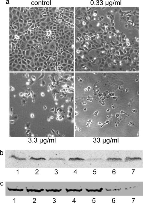
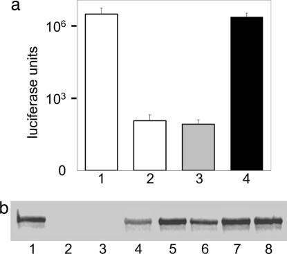
Legionella pneumophila, the causal agent of Legionnaires' disease, is an intracellular parasite and invades and proliferates within different eukaryotic cells, including human alveolar macrophages. After several 100-fold multiplication within host cells, the pathogens are released for new invasion by induction of apoptosis or necrosis. Here we report that L. pneumophila produces a glucosyltransferase, which selectively modifies an approximately 50-kDa mammalian protein by using UDP-glucose as a cosubstrate. MS analysis identified the protein substrate as the mammalian elongation factor (EF)1A. Legionella glucosyltransferase modifies its eukaryotic protein substrate at serine-53, which is located in the GTPase domain of the EF. Glucosylation of EF1A results in inhibition of eukaryotic protein synthesis and death of target cells. Our findings show a mode of inhibition of protein synthesis by microbial pathogens and offer a perspective for understanding of the host-pathogen interaction of L. pneumophila.


Conflict of interest statement

The authors declare no conflict of interest.

Figures

Fig. 1.
Fig. 1.
Glucosylation by Lgt1. (a) Glucosylation of target proteins in Caco-2 enterocyte lysate by Lgt1. Lanes 1–4 show SDS/PAGE of purified recombinant glucosyltransferases (2–4 μg per track, Coomassie staining). Lanes: 1, wild-type Lgt1; 2, D198N Lgt1; 3, D246N Lgt1; 4, D246N/D248N Lgt1. Lanes 5–10 show autoradiography of glucosylation caused by the glucosyltransferases. Caco-2 lysate was incubated with recombinant wild-type and mutant Lgt1 in the presence of UDP-[14C]glucose for 1 h. Lanes: 5, wild-type Lgt1 without Caco-2 cell extract (negative control); 6, wild-type Lgt1 plus Caco-2 cell extract; 7, D198N Lgt1 plus Caco-2 cell extract; 8, D246N Lgt1 plus Caco-2 cell extract; 9, D246N/D248N Lgt1 plus Caco-2 cell extract; 10, Caco-2 cell extract without Lgt1 (negative control). Molecular mass markers are indicated on the left. (b) Cosubstrate specificity of Lgt1. Glucosylation of Caco-2 cell lysates was performed with Lgt1 in the presence of 10 μM UDP-[14C]glucose and without (lane 1) or with (lanes 2–8) 50 μM each of unlabeled UDP-glucose (lane 2), UDP-galactose (lane 3), UDP-N-acetyl-galactosamine (lane 4), UDP-N-acetyl-glucosamine (lane 5), UDP-glucuronic acid (lane 6), GDP-mannose (lane 7), and glucose (lane 8).
Fig. 2.
Fig. 2.
Analyses of target proteins of Lgt1 by MS and mutagenesis studies. (a and b) MS analysis of proteins in Caco-2 cell lysate glucosylated by Lgt1. (a) The fragment spectrum of the nonglucosylated peptide GSF(K-dimethyl)YAWVLDK of eEF1A1. (b) The fragment spectrum of the glucosylated peptides that were only found in samples exposed to Lgt1. The peaks labeled bn (b-ions) indicate N-terminal fragment ions, and the peaks labeled yn C-terminal fragment ions. The total mass of the glucosylated peptides is precisely 162 Da higher, consistent with a covalent modification by a hexose. The fragment spectrum in b demonstrates that all fragment ions that contain the N-terminal serine (the b-ions) are shifted by 162 Da in comparison to the nonglucosylated peptide fragment spectrum in a. Each of these b-ions is accompanied by a satellite fragment marked with an asterisk, representing a loss 162 Da corresponding to a hexose group. Such an additional fragment often is seen for covalent modifications by a single hexose group. The vertical arrows indicate that ion intensity was cut for adaptation to figure size; X2 and X4 indicate multiplication of ion intensity for better presentation of data and (M+2H)2+ indicates that the doubly charged peptide had been fragmented. (c) Glucosylation of recombinant GST-tagged wild-type and mutant eEF1A1 by Lgt1. Lanes 1–3 show SDS/PAGE of purified recombinant EF1A (Coomassie staining). Lanes: 1, wild-type eEF1A1-GST; 2, eEF1A1-GST with S53T mutation; 3, eEF1A1-GST with S53A mutation. Protein load is 3–4 μg per track. Lanes 4–8 show autoradiographic analysis of substrate activity of purified recombinant EF1A. Wild-type and mutant eEF1A1-GST fusion proteins were incubated with Lgt1 for 1 h in the presence of UDP-[14C]glucose. Lanes: 4, Lgt1 without eEF1A1-GST (negative control); 5, Lgt1 plus wild-type eEF1A1-GST; 6, Lgt1 plus eEF1A1-GST with S53T mutation; 7, Lgt1 plus eEF1A1-GST with S53A mutation; 8, wild-type eEF1A1-GST without Lgt1 (negative control). The position of GST-tagged EF1A is marked on the right. Please note the weak autoglucosylation activity of Lgt1 in lanes 4–7.
Fig. 3.
Fig. 3.
Effects of Lgt1 on host cells. (a) Morphological changes in EBL cells induced by Lgt1. EBL cells were electroporated in the presence of Lgt1 at concentrations of 33, 3.3, and 0.33 μg/ml, with double D246N/D248N Lgt1 mutant at 33 μg/ml (control). Thereafter the cells were replated. After 48 h of incubation, microscopic pictures of cells were taken. (b) Back-glucosylation of the protein substrate of Lgt1 in lysate of EBL cells previously electroporated with Lgt1. Shown are samples from cell lysates electroporated with Lgt1 (3.3 μg/ml; lanes 1, 3, and 5), double D246N/D248N Lgt1 mutant (3.3 μg/ml; lanes 2, 4, and 6), or without Legionella proteins (lane 7). EBL cells were collected 30 min (lanes 1 and 2), 1.5 h (lane 3 and 4), and 4.5 h (lane 5, 6, and 7) after electroporation and used in the glucosylation assay with Lgt1 and UDP-[14C]glucose. Labeled proteins were analyzed by SDS/PAGE and phosphorimaging (shown). (c) Back-glucosylation of the protein substrate of Lgt1 in lysate of A549 cells, previously infected with L. pneumophila. A549 cells were infected with L. pneumophila at a multiplicity of infection of 10. The cells were harvested 1, 2, 3, 4, 8, and 24 h after start of the infection and lysed as described in Materials and Methods. Cell lysate was glucosylated with Lgt1 (4 μg) in the presence of UDP-[14C]glucose. Labeled proteins were analyzed by SDS/PAGE (see Fig. 6, which is published as supporting information on the PNAS web site) and phosphorimaging for [14C]glucosylation (shown). Lanes: 1, noninfected cells; 2–7, infected cells 1, 2, 3, 4, 8 and 24 h after start of the infection, respectively. Shown is a representative experiment that was repeated several times with similar results.
Fig. 4.
Fig. 4.
Inhibition of transcription/translation by Lgt1. (a) Luminescence assay. A transcription/translation reaction was performed in the absence (columns 1 and 2) or presence of recombinant wild-type (column 3) or D246N-mutated (column 4) Lgt1 (each at 2.4 μg/ml). In these experiments, matrix DNA was luciferase gene-containing plasmid (columns 1, 3, and 4). Column 2 is a negative control without matrix DNA. All measures were done in triplicate. Error bars indicate standard deviations. Differences in the values of columns 1 and 2 with those of columns 3 and 4 are statistically significant (P < 0.01). White columns show control results, the gray column shows wild-type Lgt1, and the black column shows mutated Lgt1. The y axis represents logarithmic scale. (b) Autoradiographic assay. A transcription/translation reaction has been performed in the absence (lanes 1 and 2) or presence (lanes 3–8) of recombinant wild-type (lanes 3–5) or D246N-mutated (lanes 6–8) Lgt1. Concentrations of L. pneumophila proteins were 2.4 μg/ml in lanes 3 and 6; 240 ng/ml in lanes 4 and 7; and 24 ng/ml in lanes 5 and 8. Matrix DNA present in lanes 1 and 3–8 was luciferase gene-containing plasmid coding for an ≈60-kDa protein. Lane 2 is a negative control without matrix DNA.
Fig. 5.
Fig. 5.
Inhibition of [35S]methionine incorporation by Lgt1. EBL cells were electroporated with wild-type (columns 2, 4, 6, and 8) and double D246N/D248N Lgt1 mutant (columns 3, 5, 7, and 9) in methionine-free MEM. Two hours after intoxication, cells were pulsed with 0.5 μCi [35S]methionine for a further 3 h, lysed, and assayed for incorporation of radioactivity into proteins. Columns: 1, control cells electroporated without Legionella proteins; 2–9, cells were electroporated with Legionella proteins at 33 ng/ml (columns 2 and 3), 0.33 μg/ml (columns 4 and 5), 3.3 μg/ml (columns 6 and 7), or 33 μg/ml (columns 8 and 9). All measures have been done in triplicate. Error bars indicate standard deviations. Differences between the values in columns 6 and 7 with those in columns 8 and 9 are statistically significant (P < 0.01). The white column represents the control experiment, the gray columns represent wild-type Lgt1, and black columns represent mutant Lgt1.

Similar articles

Cited by

References

    1. Fields BS, Benson RF, Besser RE. Clin Microbiol Rev. 2002;15:506–526. - PMC - PubMed
    1. Marston BJ, Plouffe JF, File TM, Jr, Hackman BA, Salstrom SJ, Lipman HB, Kolczak MS, Breiman RF. Arch Intern Med. 1997;157:1709–1718. - PubMed
    1. Horwitz MA. J Exp Med. 1983;158:2108–2126. - PMC - PubMed
    1. Horwitz MA, Maxfield FR. J Cell Biol. 1984;99:1936–1943. - PMC - PubMed
    1. Summersgill JT, Raff MJ, Miller RD. Microb Pathog. 1988;5:41–47. - PubMed

Publication types

MeSH terms

Substances

LinkOut - more resources