. 2007 Feb 15;109(4):1345-52.
doi: 10.1182/blood-2006-07-037952. Epub 2006 Oct 26.

Orientation of endothelial cell division is regulated by VEGF signaling during blood vessel formation

Affiliations

Orientation of endothelial cell division is regulated by VEGF signaling during blood vessel formation

Gefei Zeng et al. Blood. .

Abstract

New blood vessel formation requires the coordination of endothelial cell division and the morphogenetic movements of vessel expansion, but it is not known how this integration occurs. Here, we show that endothelial cells regulate division orientation during the earliest stages of blood vessel formation, in response to morphogenetic cues. In embryonic stem (ES) cell-derived vessels that do not experience flow, the plane of endothelial cytokinesis was oriented perpendicular to the vessel long axis. We also demonstrated regulated cleavage orientation in vivo, in flow-exposed forming retinal vessels. Daughter nuclei moved away from the cleavage plane after division, suggesting that regulation of endothelial division orientation effectively extends vessel length in these developing vascular beds. A gain-of-function mutation in VEGF signaling increased randomization of endothelial division orientation, and this effect was rescued by a transgene, indicating that regulation of division orientation is a novel mechanism whereby VEGF signaling affects vessel morphogenesis. Thus, our findings show that endothelial cell division and morphogenesis are integrated in developing vessels by flow-independent mechanisms that involve VEGF signaling, and this cross talk is likely to be critical to proper vessel morphogenesis.


Figures

Figure 1
Figure 1
Regulated endothelial cell division orientation may affect vessel morphogenesis: a model. This model shows how endothelial cell divisions whose orientation is regulated relative to the long axis of the vessel could affect vessel shape. Endothelial divisions oriented perpendicular to the vessel long axis (blue vessel on top) would effectively lengthen the vessel, whereas divisions oriented parallel to the vessel long axis (yellow vessel on bottom) would effectively increase the vessel diameter. Microtubules and spindles are shown in red, and DNA/chromosomes are brown.
Figure 2
Figure 2
Endothelial cell divisions are oriented perpendicular to the vessel long axis in ES cell–derived vessels. Mouse ES cells (WT; Tg PECAM-H2B-GFP) differentiated to day 7 to 8 were imaged for several hours prior to fixation and staining for PECAM-1. (A) A confocal image showing H2B-GFP signal in green (i), the same field with PECAM-1 stain in red after fixation (ii), and the overlay of the 2 images (iii), showing the H2B-GFP signal and PECAM-1 stain in the same cells. (B) The top portion of the images in panel A, showing time lapse images from 0 minute (i) to 292 minutes (vi). Panel iv was used to calculate the angle of division relative to the vessel axis, according to the drawn yellow lines. The numbers in the lower right represent elapsed time in minutes. (C) Calculation of division angles relative to the vessel long axis. Top panel was colorized as in Figure 1, except the chromosomes are blue. The 90-degree division angle is perpendicular to the vessel long axis, and the 0-degree division angle is parallel to the vessel long axis; n = 125 divisions. (D) Representation of endothelial division angles, with the vessel long axis diagrammed by the long horizontal lines. Each shorter line represents 3 angle measurements that were close or equivalent to each other. For a video of panel B, see Video S1 (available on the Blood website; see the Supplemental Videos link at the top of the online article).
Figure 3
Figure 3
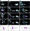
Daughter cells maintain their division orientation as they migrate after most divisions. ES cell–derived vessels (WT; Tg PECAM H2B-GFP) were imaged, and after divisions were scored (i panels) the 2 daughter nuclei (pink) were followed for 1 to 2 hours further. The numbers in the lower right represent elapsed time in minutes. In all cases ii panels show the final image scored. The iii panels diagram the movement of each daughter nucleus (shown in pink) after division: one daughter nucleus is tracked with a green line and the other with a pink line. The vessel is shown by black lines, the vessel long axis is shown by the broken blue line, and the division angle is shown by the broken orange line. (A) A division perpendicular to the vessel long axis, where the daughter cells maintained the division orientation after 1 hour (n = 61 of 61). (B) A division parallel to the vessel long axis, where the daughter cells maintained the division orientation after 1 hour (n = 7 of 10). (C) A division parallel to the vessel long axis, where the daughter cells changed position relative to the division orientation after 102 minutes (n = 3 of 10).
Figure 4
Figure 4
Endothelial cell divisions orient perpendicular to the nearest long axis. ES cell–derived vessels (WT; Tg PECAM-eGFP) were imaged for various lengths of time, and divisions in or around sprouts were scored for division orientation. In all series, the yellow cell is the scored endothelial cell just prior to division, the pink cells are the daughter cells, and the numbers in the lower right represent elapsed time in minutes. In all cases panel v diagrams the division that was scored, with the parent vessel long axis the broken blue line, the sprout long axis the dotted green line, and the division angle the broken orange line. (A) An endothelial division that occurred prior to nearby sprout formation. Although the sprout (arrows in panels iii and iv) migrated almost perpendicular to the vessel long axis, the division was oriented perpendicular to the parent vessel long axis (n = 2 of 2). (B) An endothelial division that occurred in the sprout field, at the base of a formed sprout but in the parent vessel. These divisions also oriented perpendicular to the parent vessel long axis (n = 6 of 6). (C) In contrast, an endothelial division that occurred within a formed sprout oriented perpendicular to the sprout vessel long axis (n = 5 of 5).
Figure 5
Figure 5
Endothelial cell divisions are oriented perpendicular to the vessel long axis in retinal vessels in vivo. Rat retinas were harvested on days P3 to P5 and processed for staining as described with Griffonia B4 isolectin (green) to visualize vessels and phosphohistone H3 (red) to visualize DNA in mitotic cells. (A) Several examples of divisions in retinal vessels that were scored for division angle. (B) Calculation of division angles relative to vessel long axis. Ninety degrees is perpendicular to the vessel long axis, and 0 degree is parallel to the vessel long axis; n = 86 divisions. (C) Representation of endothelial division angles, with the vessel long axis diagrammed by the long horizontal lines. Each shorter line represents 3 angle measurements that were close or equivalent to each other.
Figure 6
Figure 6
Endothelial cell divisions are randomly oriented flt-1−/− ES cell–derived vessels. Mouse ES cells (flt-1−/−; Tg PECAM-H2B-GFP) differentiated to day 7 to 8 were imaged for several hours prior to fixation and staining for PECAM-1. (A) Time lapse images of a representative movie from 0 minute (i) to 240 minutes (vi), showing the H2B signal in vessels. Panel iii was used to calculate the angle of division relative to the vessel axis, according to the drawn yellow lines. The numbers in the lower right represent elapsed time in minutes. (B) Calculation of division angles relative to the vessel long axis. WT endothelial division angles are shown in blue, and flt-1−/− endothelial division angles are shown in purple. Ninety degrees is perpendicular to the vessel long axis, and 0 degree is parallel to the vessel long axis; n = 125 divisions for WT vessels (same data as shown in Figure 2) and n = 93 divisions for flt-1−/− vessels. (C) Representation of endothelial division angles, with the vessel long axis diagrammed by the long horizontal lines. Each shorter line represents 3 angle measurements that were close or equivalent to each other. For the video of panel A, see Video S2.
Figure 7
Figure 7
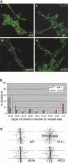
Randomized endothelial division orientation is rescued by a sflt-1 transgene that rescues flt-1−/− vessel dysmorphogenesis. ES cell–derived vessels were wild-type (WT), flt-1−/−, or flt-1−/− with a sflt-1 transgene (flt-1−/−;Tg PECAM sflt-1#26, indicated as sflt26; or flt-1−/−;Tg PECAM sflt-1#33, as sflt33). Cultures were differentiated to day 8, fixed, and stained for PECAM-1 (green) and phosphohistone H3 (red). (A) Representative vessels of the indicated genotypes, with white arrows pointing to endothelial divisions that were scored. (B) Graphic representation of division angles from the different genetic backgrounds. Ninety degrees is perpendicular to the vessel long axis, and 0 degree is parallel to the vessel long axis. WT is blue (n = 19), flt-1−/− is purple (n = 29), sflt26 is yellow (n = 25), and sflt33 is light green (n = 23). (C) Representation of endothelial division angles, with the vessel long axis diagrammed by the long horizontal lines. Each shorter line represents a single angle measurement.

Similar articles

Cited by

References

    1. Risau W. Mechanisms of angiogenesis. Nature. 1997;386:671–674. - PubMed
    1. Eichmann A, Makinen T, Alitalo K. Neural guidance molecules regulate vascular remodeling and vessel navigation. Genes Dev. 2005;19:1013–1021. - PubMed
    1. Coultas L, Chawengsaksophak K, Rossant J. Endothelial cells and VEGF in vascular development. Nature. 2005;438:937–945. - PubMed
    1. Rousseau S, Houle F, Huot J. Integrating the VEGF signals leading to actin-based motility in vascular endothelial cells. Trends Cardiovasc Med. 2000;10:321–327. - PubMed
    1. Kliche S, Waltenberger J. VEGF receptor signaling and endothelial function. IUBMB Life. 2001;52:61–66. - PubMed

Publication types

Substances