. 2006 Oct;2(10):e177.
doi: 10.1371/journal.pgen.0020177. Epub 2006 Sep 1.

RanBP2 modulates Cox11 and hexokinase I activities and haploinsufficiency of RanBP2 causes deficits in glucose metabolism

Affiliations

RanBP2 modulates Cox11 and hexokinase I activities and haploinsufficiency of RanBP2 causes deficits in glucose metabolism

Azamat Aslanukov et al. PLoS Genet. 2006 Oct.

Abstract

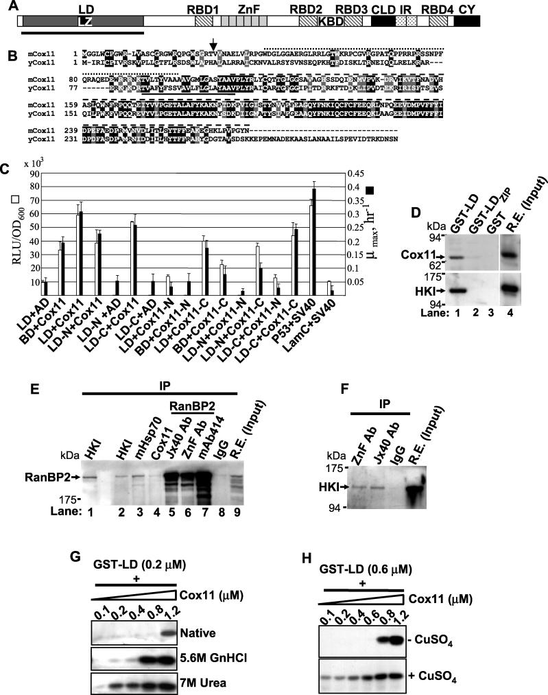
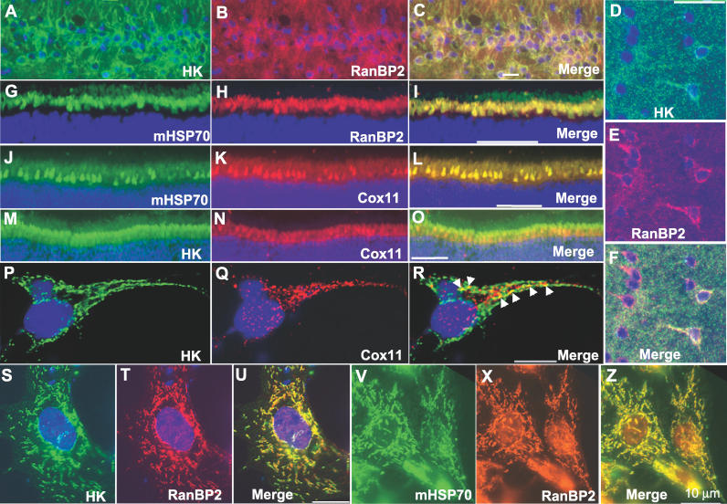
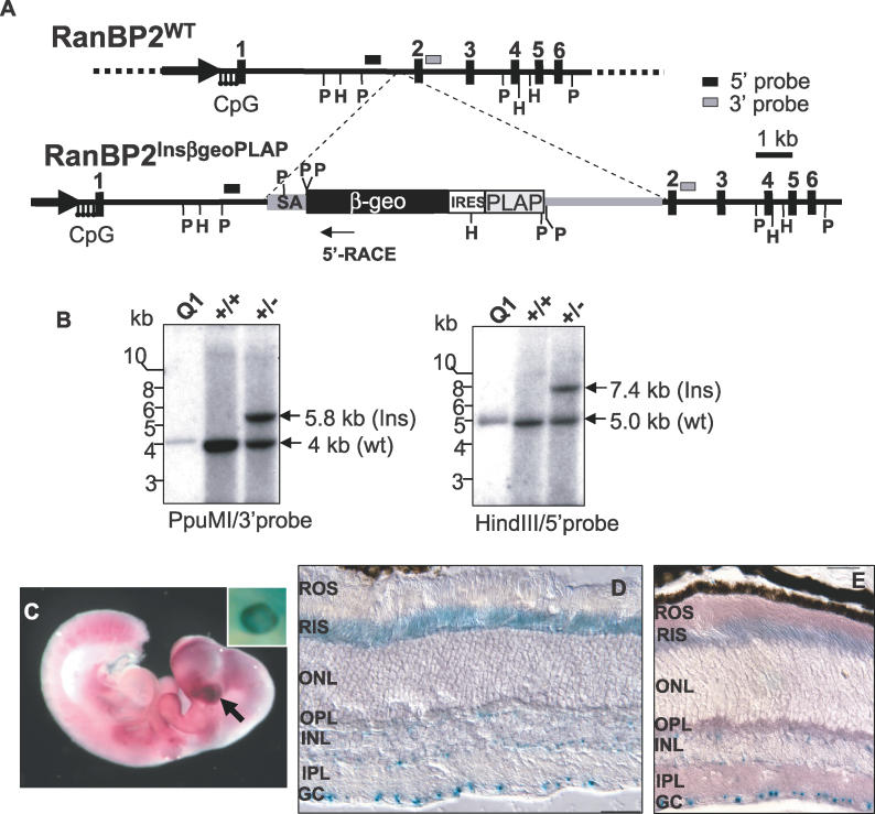
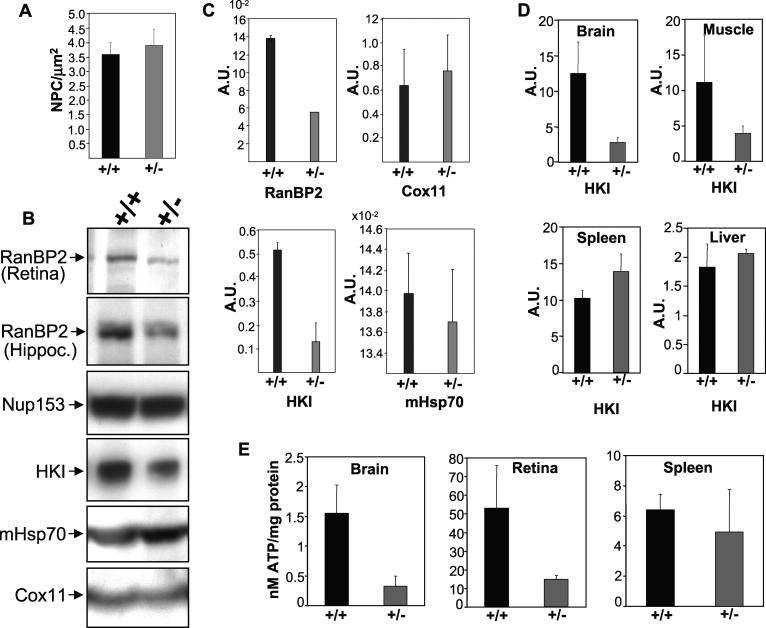
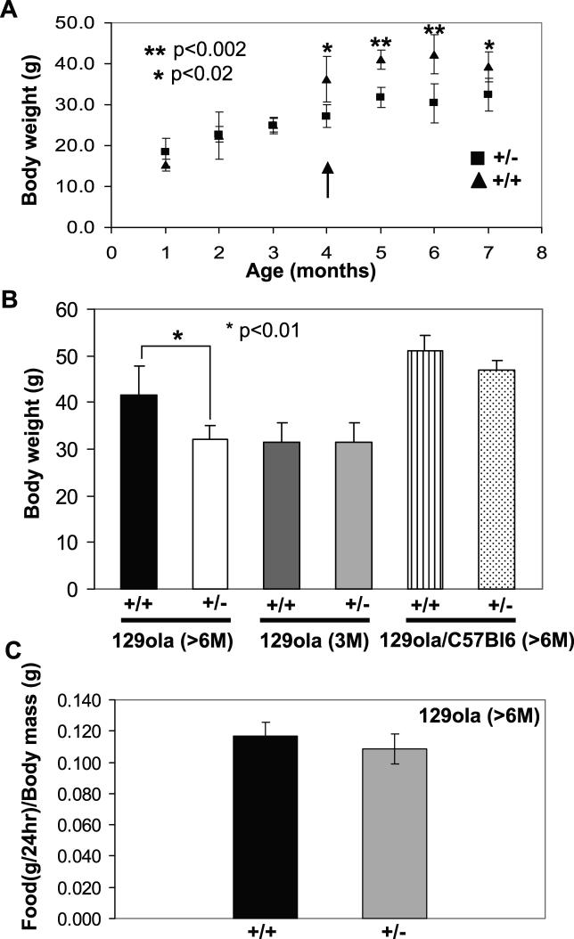
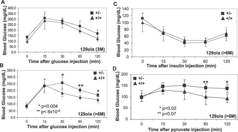
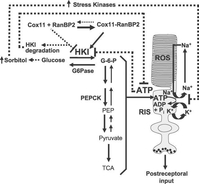
The Ran-binding protein 2 (RanBP2) is a large multimodular and pleiotropic protein. Several molecular partners with distinct functions interacting specifically with selective modules of RanBP2 have been identified. Yet, the significance of these interactions with RanBP2 and the genetic and physiological role(s) of RanBP2 in a whole-animal model remain elusive. Here, we report the identification of two novel partners of RanBP2 and a novel physiological role of RanBP2 in a mouse model. RanBP2 associates in vitro and in vivo and colocalizes with the mitochondrial metallochaperone, Cox11, and the pacemaker of glycolysis, hexokinase type I (HKI) via its leucine-rich domain. The leucine-rich domain of RanBP2 also exhibits strong chaperone activity toward intermediate and mature folding species of Cox11 supporting a chaperone role of RanBP2 in the cytosol during Cox11 biogenesis. Cox11 partially colocalizes with HKI, thus supporting additional and distinct roles in cell function. Cox11 is a strong inhibitor of HKI, and RanBP2 suppresses the inhibitory activity of Cox11 over HKI. To probe the physiological role of RanBP2 and its role in HKI function, a mouse model harboring a genetically disrupted RanBP2 locus was generated. RanBP2(-/-) are embryonically lethal, and haploinsufficiency of RanBP2 in an inbred strain causes a pronounced decrease of HKI and ATP levels selectively in the central nervous system. Inbred RanBP2(+/-) mice also exhibit deficits in growth rates and glucose catabolism without impairment of glucose uptake and gluconeogenesis. These phenotypes are accompanied by a decrease in the electrophysiological responses of photosensory and postreceptoral neurons. Hence, RanBP2 and its partners emerge as critical modulators of neuronal HKI, glucose catabolism, energy homeostasis, and targets for metabolic, aging disorders and allied neuropathies.


Conflict of interest statement

Competing interests. The authors have declared that no competing interests exist.

Figures

Figure 1
Figure 1. The LD of RanBP2 Interacts with Cox11 and HKI
(A) Primary structure of RanBP2 and its structural/functional domains. The N-terminal LD of RanBP2 is underlined. (B) Sequence alignment of murine and yeast Cox11. The yeast Cox11 C- and N-terminal domains are poorly conserved. Arrow and solid line denote the predicted mitochondrial cleavage site and membrane-spanning domain. The dotted and dashed lines above the aligned sequences represent, respectively, Cox11-N and Cox11-C constructs shown in Figure 1C. (C) Structure-function analysis of the interaction between the LD of RanBP2 and Cox11. Optimal interaction between the LD and Cox11 occurred in the presence of constructs comprising both the complete LD and Cox11. Although removal of the cytosolic N-terminal (Cox11-C) significantly decreased the interaction with LD, the mitochondrial intermembrane domain of Cox11 (Cox11-C) together with the C-terminal half of LD (LD-C) retained most of the interaction activity. LD-N and LD-C ended and began with the leucine zipper domain of RanBP2. White and black bars denote β-galactosidase activity and growth rates in selective growth medium, respectively. Results shown represent the mean ± SD, n = 3. (D) GST pull-down assays with the LD of RanBP2 and its leucine zipper domain and retinal extracts. The LD, but not the leucine zipper domain of RanBP2, associate with Cox11 (top panel, lane 1) and HKI (bottom panel, lane 1). (E) Coimmunoprecipitation of RanBP2 with antibodies against its molecular partners shows that RanBP2 forms a complex in vivo with HKI (lanes 1 and 2), mHsp70 (lane 3), and Cox11 (lane 4). Lanes 5, 6, and 7 are control immunoprecipitation reactions with different antibodies against the RanBP2 domains, KBD, ZnF, and XAFXFG of nucleoporins. (F) Reciprocal coimmunoprecipitation of HKI with antibodies against RanBP2 (used and shown in (E)). (G) Reconstitution pull-down assays with purified LD and increasing concentrations of native (top panel), denatured (middle panel), and partially denatured (bottom panel) Cox11, respectively, in the absence and presence of denaturating agent, GnHCl and chaotropic agent, urea. Folding intermediates (lower panel) of Cox11 exhibit the highest binding activity toward the LD of RanBP2. (H) Similar experiments as in (G) but in the presence of native Cox11 expressed in the absence (top panel) or presence (bottom panel) of CuSO4. The mature isoform of the metallochaperone has an increased affinity toward the LD of RanBP2. LD, leucine-rich domain; LZ, leucine zipper domain; RBD1–4, Ran-binding domains 1–4; ZnF, zinc finger cluster domain; KBD, kinesin (KIF5B/KIF5C)-binding domain; CLD, cyclophilin-like domain; IR, internal repeat domain; CY, cyclophilin domain.
Figure 2
Figure 2. Effect of Cox11 and RanBP2 on HKI Activity
(A) Saturation kinetics, rate versus glucose of HKI (0.24 μg) in the absence (solid circles) and presence of Cox11 (open circles, 0.25 nM; solid triangles, 7.5 nM). The activity of HKI decreases with increasing concentrations of Cox11. No measurable HKI activity was recorded in the presence of 15 nM of Cox11 (unpublished data). (B) Hanes-Wolf plot of (A) (1/rate versus glucose) in the absence and presence of fixed concentrations of Cox11. Linearity of reciprocal plots also supported the hyperbolic behavior of the reactions (unpublished data). Cox11 behaves as a noncompetitive inhibitor of HKI by reducing the V max of HKI but not its K m toward glucose. (C) HKI rate is plotted as a function of LD concentration at saturating glucose and fixed Cox11 (7.5 nM) concentrations. Note that increasing concentrations of the LD of RanBP2 reverse the inhibition of HKI activity by Cox11. A half-maximal effect of the LD of RanBP2 on HKI activity in the presence of 7.5 nM of Cox11 was observed at a concentration of ~0.05 nM of LD. (D) Rate versus glucose plot in the absence and presence of the LD of RanBP2. At a saturating concentration of the LD of RanBP2 (3.75 nM), the HKI activity was reduced by about 20%. v, rate; S, glucose.
Figure 3
Figure 3. Localization of RanBP2 and Its LD Molecular Partners
(A–F) are thin cryosections of an area of the hipocampus (CA1 neurons) and cerebral cortex, respectively, immunostained against HKI (A and D), RanBP2 (B and E), and merged images thereof (C and F). Note that while RanBP2 and HKI are widely expressed among and colocalize to hippocampal neurons (C), HKI expression and localization with RanBP2 is restricted to a subset of cortical neurons (likely interneurons) (F). Images of the distal region of bovine retinal cryosections comprising part of the nuclear layer of photoreceptor neurons and their inner (myoid and ellipsoid) segment compartment (G–O) are immunostained against mHsp70 (G) and RanBP2 (H), mHsp70 (J) and Cox11 (K), HKI (M) and Cox11 (N), and merged images thereof (I–O). Note the prominent localization of RanBP2, mHsp70, and Cox11 at the mitochondria-rich ellipsoid compartment of photoreceptors and the colocalization of RanBP2 and Cox11 with mHsp70 (I and L), while HKI colocalization with Cox11 was limited to restricted foci (R, arrowheads). High-resolution images of dissociated primary cerebral neurons and glial cells confirmed that the colocalization of HKI and Cox11 was highly restricted (P–R), while RanBP2 extensively colocalized with HKI (S–U) and mHsp70 (V–Z). Scale bars in A–O and P–Z are 40 and 10 μm, respectively. ONL, outer nuclear layer.
Figure 4
Figure 4. Insertion Mutagenesis of the Murine RanBP2 Gene
(A) Diagram of the genomic region of RanBP2 disrupted by insertion trap mutagenesis with a bicistronic reporter vector between exon 1 and 2. The bicistronic transcript produces two proteins under regulation of RanBP2. Upon splicing of RanBP2, a fusion between exon 1 and β-geo (a fusion between the β-gal and neo genes) is generated, while human placental alkaline phophatase (PLAP) is independently translated using the internal ribosome entry site. Consistent with previous studies, the expression of the former is directed to cell bodies, while expression of the latter is targeted to the axonal processes [67,68]. Transcriptional 5′ RACE analysis detects a fusion between exon 1 and β-geo. (B) Southern analysis of the RanBP2 locus of wild-type and heterozygous genomic DNA of tails of F1 mice digested with PpuMI (left panel) and HindIII (right panel) with probes at the 3′ (left panel) and 5′ (right panel) flanking regions of the insertion breakpoint. Q1 is a cosmid containing the RanBP2 gene up to exon 20 [4]. (C) Lateroventral view of a whole-mount stain of a ~12.5 dpc heterozygous embryo for PLAP and β-gal (inset picture) activities. Although PLAP was broadly expressed (e.g., somites, limbs, and CNS), the PLAP and β-Gal (inset picture) expression was particularly high in the optic vesicle (arrow). X-gal single (D) and combined staining with PLAP (E) of a retinal section of a 3-mo-old RanBP2+/− mouse. Consistent with previous immunocytochemistry studies, β-Gal activity is detected in the neuroretinal bodies and inner segment compartment of photoreceptors with conspicuously strong expression in ganglion cells. PLAP expression is found throughout the plexiform/synaptic layers and outer segment of photoreceptors (E). GC, ganglion cell; PLAP, human placental alkaline phophatase; ROS, rod outer segment; RIS, rod inner segment; ONL, outer nuclear layer; OPL, outer plexiform (synaptic) layer; INL, inner nuclear layer; IPL, inner plexiform (synaptic) layer; GC, ganglion cell layer.
Figure 5
Figure 5. Haploinsufficiency of RanBP2 Causes a Decrease in HKI Protein and ATP Levels
(A) Quantitative analysis of NPCs in dissociated hippocampal neurons of wild-type (+/+) and heterozygote (+/−) mice upon immunostaining with mAb414. No difference in the density of NPCs (3–4 NPC/μm2) at the nuclear envelope was found between RanBP2+/+ and RanBP2+/− mice. (B) Immunoblots with anti-RanBP2/Nup153/Nup62 (mAb414), −HKI, −mHsp70, and −Cox11 antibodies of retinal (top panel) and hippocampal homogenates of +/+ and +/− mice. In comparison to RanBP2+/+, RanBP2+/− mice exhibit a reduction in the expression levels of RanBP2 and HKI but not of other proteins. (C) Quantitative analysis of relative protein expression levels of RanBP2, Cox11, HKI, and mHsp70 in the hippocampus of RanBP2+/+ and RanBP2+/−mice. There is ~2- and 4-fold reduction of RanBP2 and HKI in heterozygote mice. (D) The level of HKI is reduced in the brain but not in other non-neuronal tissues tested (muscle, spleen, and liver). (E) The total ATP level is reduced in the CNS tissues (brain and retina) but not in non-neuronal tissues tested (e.g., spleen).
Figure 6
Figure 6. RanBP2+/− Mice on High-Fat Diet Exhibit Deficits in Growth
(A) In comparison to wild-type mice, RanBP2+/− mice show slower growth rates beginning at 4 mo of age (arrow), and the difference in body weight between these is maintained afterward. Note that RanBP2+/− mice lack the growth spur observed in wild-type mice between 3 and 4 mo of age. (B) In comparison to inbred RanBP2+/−mice (129Ola genetic background), the difference in body weight between RanBP2+/+ and RanBP2+/− mice is masked upon placing these on a mixed 129Ola/C57Bl6 genetic background. (C) RanBP2+/+ and RanBP2+/−inbred mice exhibit similar rates of food consumption. Mice in (A), (B), and (C) were placed on a high-fat diet since birth (n = 5).
Figure 7
Figure 7. Metabolic Phenotypes of RanBP2+/− Inbred Mice on High-Fat Diet
(A) 3-mo-old inbred RanBP2+/− mice (n = 5) have normal glucose clearance rates upon glucose challenge and overnight fasting. (B) In contrast, 6-mo-old inbred RanBP2+/− mice (n = 5) have significantly decreased glucose clearance rates upon glucose challenge and overnight fasting. (C) Fasted 6- to 8-mo-old RanBP2+/+ and RanBP2+/− mice have no difference in insulin-mediated glucose uptake as assayed by insulin tolerance test (n = 5). (D) Pyruvate tolerance test shows normal rise in glucose but decreased glucose clearance between inbred RanBP2+/+ and RanBP2+/− mice (n = 5).
Figure 8
Figure 8. Electroretinograms from 6-Mo-Old RanBP2+/−and RanBP2+/+ Inbred Mice Showing Photoreceptor and Postreceptor Neuron Electrophysiological Response Phenotypes
(A) Scotopic (dark-adapted) responses from RanBP2+/− mice to light stimuli of increasing intensity, beginning at threshold, have reduced amplitudes compared to those observed in RanBP2+/+ mice. The three lower intensities represent responses generated in the rod photoreceptor neuronal pathway. The upper intensities are comprised of responses generated in both the rod and cone pathways. (B) Photopic (light-adapted, cone photoreceptor pathway) responses of RanBP2+/− mice to increasing light stimulus intensities also exhibited reduced amplitudes compared to those observed in RanBP2+/+ mice. (C) Average ± SE (n = 9) scotopic b-wave amplitudes from RanBP2+/− (open circles) and RanBP2+/+ (filled squares) mice representing postreceptoral neuron function. (Note: log amplitude scale.) (D) Average ± SE (n = 5) scotopic a-wave amplitudes, representing photoreceptor function, for RanBP2+/− and RanBP2+/+ mice in response to bright flashes. Amplitudes of responses from RanBP2+/− mice were lower over the entire range of stimulus intensities for both b- and a-waves. Asterisks represent significant differences between the groups (Student's t test, p < 0.05). Statistical significance was found across all intensities for b-wave amplitudes (2-way ANOVA, p < 0.0001), but not for the a-wave.
Figure 9
Figure 9. Model Depicting a Role of RanBP2 and Some of Its Partners in Metabolic and Neuronal Function
RanBP2 interacts with Cox11 and HKI and the triad is in equilibrium under normal physiological conditions. RanBP2 prevents the inhibition of HKI by Cox11 and its degradation. The ultimate effect of RanBP2 on its partners is the stimulation of the glycolytic pathway and production of ATP. The glycolytic pathway is critical to fuel the constitutive Na+/K+-ATPase pump to maintain the dark current between the inner and outer segment compartments of photosensory neurons. A deficit (haploinsufficiency) in RanBP2 disturbs the equilibrium between RanBP2, HKI, and Cox11. This pathophysiological event promotes the destabilization and degradation of HKI and a decrease in ATP production required to maintain the depolarization state neurons, and, hence, a reduction in the response of receptoral and postreceptoral neurons. A reduction in ATP levels also negatively modulates HKI activity/level. Decreased levels of HKI promote intracellular hyperglycemia and activate stress kinases, which modulate negatively the Na+/K+-ATPase pump by phosphorylation. Pathophysiological pathways promoted by RanBP2 haploinsufficiency are represented by dash lines. RIS, rod inner segment; ROS, rod outer segment.

References

    1. Ferreira PA, Hom JT, Pak WL. Retina-specifically expressed novel subtypes of bovine cyclophilin. J Biol Chem. 1995;270:23179–23188. - PubMed
    1. Wu J, Matunis MJ, Kraemer D, Blobel G, Coutavas E. Nup358, a cytoplasmically exposed nucleoporin with peptide repeats, Ran-GTP binding sites, zinc fingers, a cyclophilin A homologous domain, and a leucine-rich region. J Biol Chem. 1995;270:14209–14213. - PubMed
    1. Yokoyama N, Hayashi N, Seki T, Pante N, Ohba T, et al. A giant nucleopore protein that binds Ran/TC4. Nature. 1995;376:184–188. - PubMed
    1. Fauser S, Aslanukov A, Roepman R, Ferreira PA. Genomic organization, expression, and localization of murine Ran-binding protein 2 (RanBP2) gene. Mamm Genome. 2001;12:406–415. - PubMed
    1. Bernad R, van der Velde H, Fornerod M, Pickersgill H. Nup358/RanBP2 attaches to the nuclear pore complex via association with Nup88 and Nup214/CAN and plays a supporting role in CRM1-mediated nuclear protein export. Mol Cell Biol. 2004;24:2373–2384. - PMC - PubMed

Publication types

MeSH terms