. 2006 Oct;15(5):657-86.
doi: 10.1111/j.1365-2583.2006.00689.x.

Characteristics of the nuclear (18S, 5.8S, 28S and 5S) and mitochondrial (12S and 16S) rRNA genes of Apis mellifera (Insecta: Hymenoptera): structure, organization, and retrotransposable elements

Affiliations

Characteristics of the nuclear (18S, 5.8S, 28S and 5S) and mitochondrial (12S and 16S) rRNA genes of Apis mellifera (Insecta: Hymenoptera): structure, organization, and retrotransposable elements

J J Gillespie et al. Insect Mol Biol. 2006 Oct.

Abstract

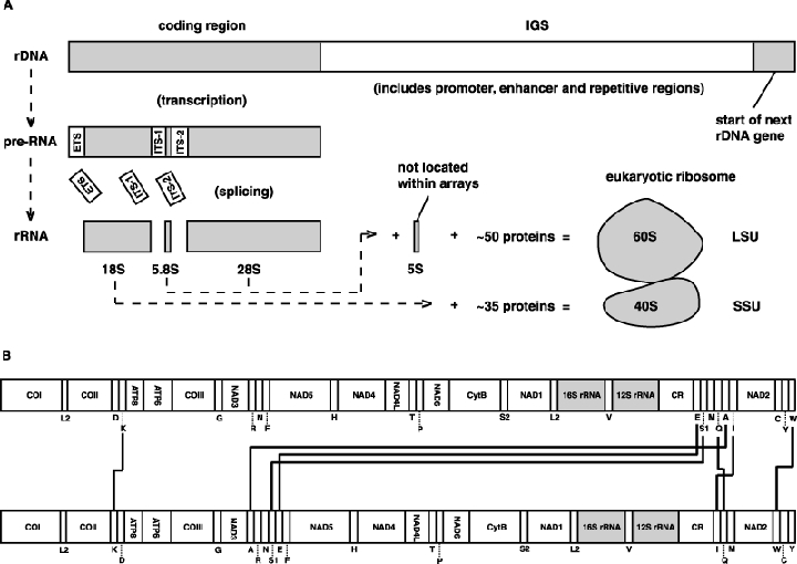
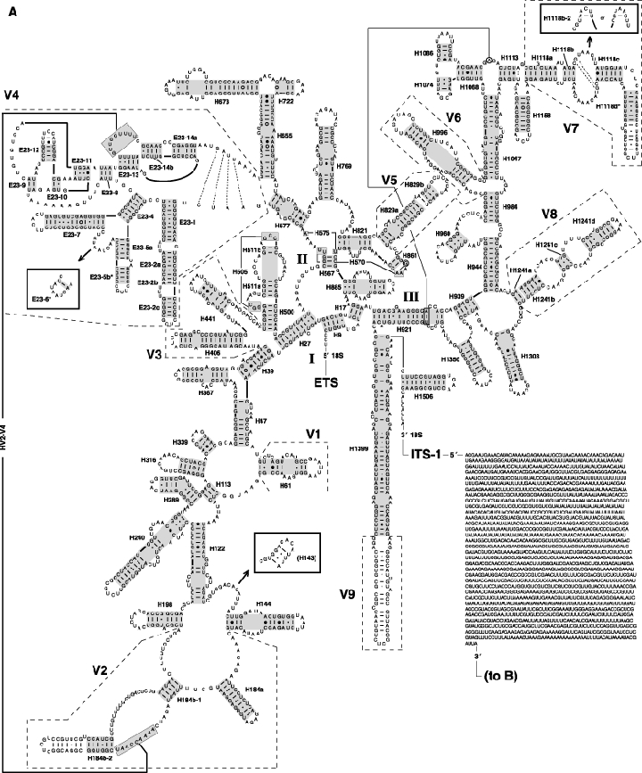
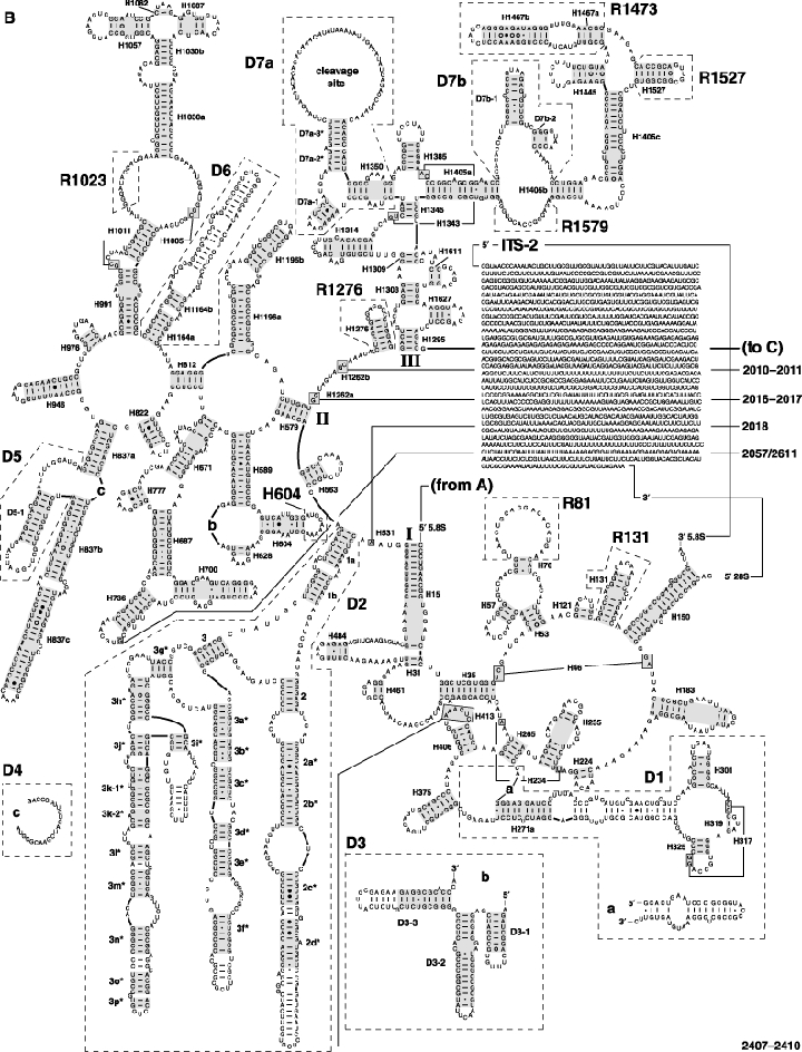
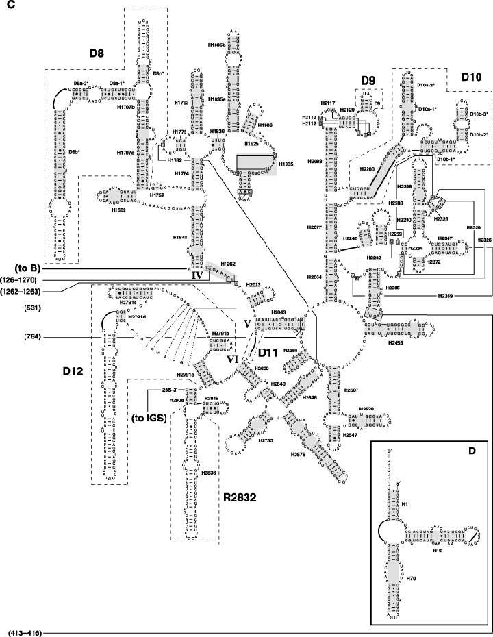
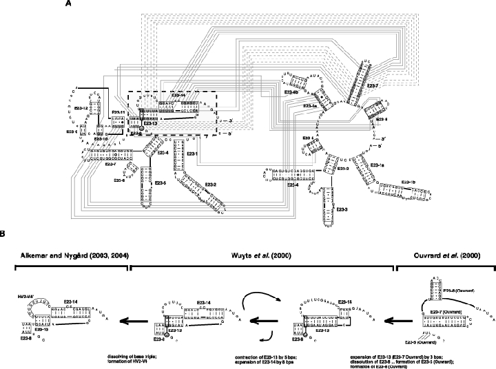
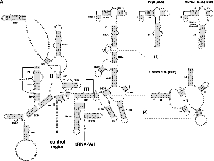
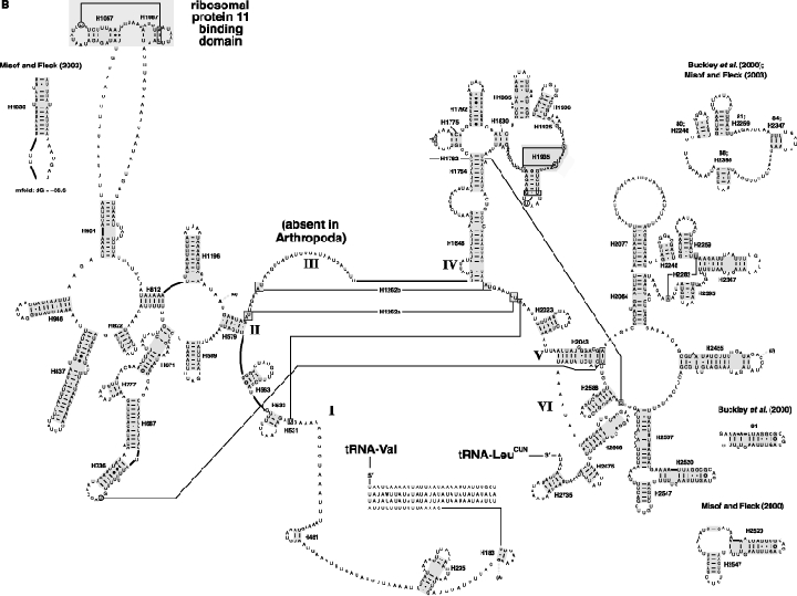
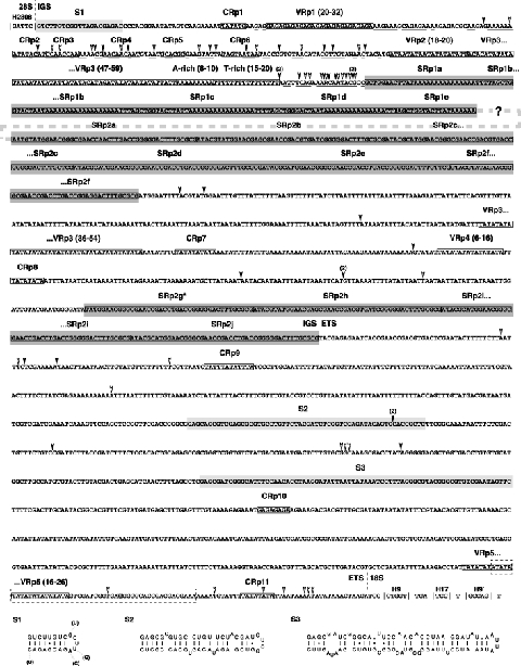
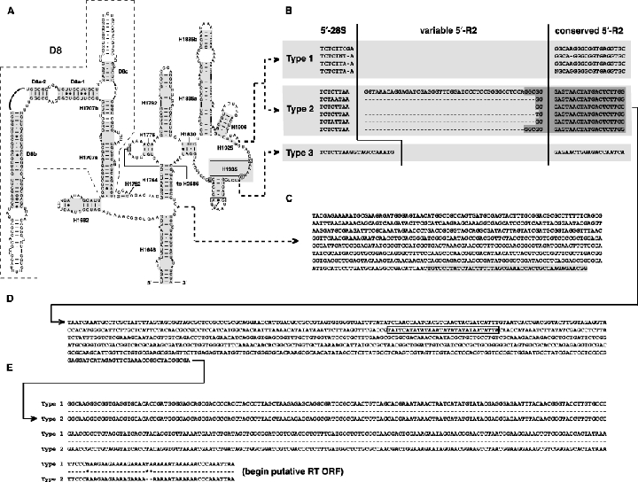
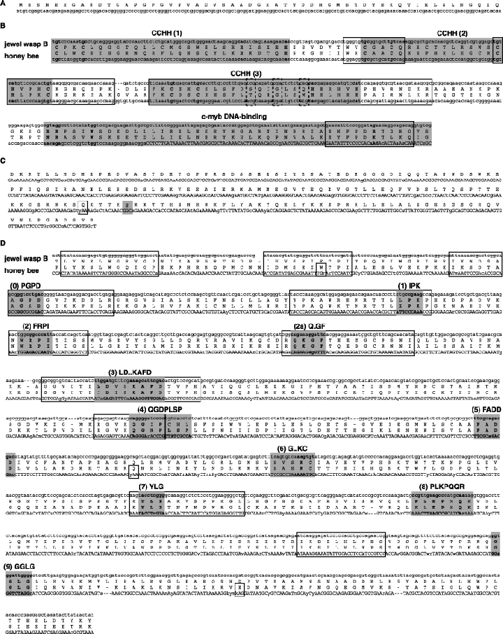
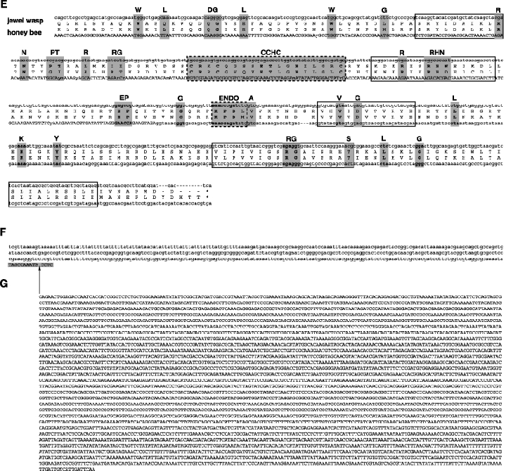
As an accompanying manuscript to the release of the honey bee genome, we report the entire sequence of the nuclear (18S, 5.8S, 28S and 5S) and mitochondrial (12S and 16S) ribosomal RNA (rRNA)-encoding gene sequences (rDNA) and related internally and externally transcribed spacer regions of Apis mellifera (Insecta: Hymenoptera: Apocrita). Additionally, we predict secondary structures for the mature rRNA molecules based on comparative sequence analyses with other arthropod taxa and reference to recently published crystal structures of the ribosome. In general, the structures of honey bee rRNAs are in agreement with previously predicted rRNA models from other arthropods in core regions of the rRNA, with little additional expansion in non-conserved regions. Our multiple sequence alignments are made available on several public databases and provide a preliminary establishment of a global structural model of all rRNAs from the insects. Additionally, we provide conserved stretches of sequences flanking the rDNA cistrons that comprise the externally transcribed spacer regions (ETS) and part of the intergenic spacer region (IGS), including several repetitive motifs. Finally, we report the occurrence of retrotransposition in the nuclear large subunit rDNA, as R2 elements are present in the usual insertion points found in other arthropods. Interestingly, functional R1 elements usually present in the genomes of insects were not detected in the honey bee rRNA genes. The reverse transcriptase products of the R2 elements are deduced from their putative open reading frames and structurally aligned with those from another hymenopteran insect, the jewel wasp Nasonia (Pteromalidae). Stretches of conserved amino acids shared between Apis and Nasonia are illustrated and serve as potential sites for primer design, as target amplicons within these R2 elements may serve as novel phylogenetic markers for Hymenoptera. Given the impending completion of the sequencing of the Nasonia genome, we expect our report eventually to shed light on the evolution of the hymenopteran genome within higher insects, particularly regarding the relative maintenance of conserved rDNA genes, related variable spacer regions and retrotransposable elements.


Figures

Figure 1
Figure 1
Organization of the rRNA genes within the honey bee genome. (A) Typical organization of the nuclear rRNA genes of eukaryotes. IGS, intergenic spacer; SSU, small subunit; LSU, large subunit. (B) Position of the rRNA genes (shaded) within the honey bee mitochondrial genome (top) and the ancestral arthropod mitochondrial genome (bottom). Rearrangements in the honey bee genome are depicted with solid lines. See Clary & Wolstenholme (1985, 1987) and Crozier & Crozier (1993) for information on honey bee mitochondrial genes.
Figure 2
Figure 2
The secondary structure model of the nuclear rRNA (18S + 5.8S + 28S + 5S) from the honey bee, Apis mellifera. Variable regions are enclosed within dashed boxes and the naming follows either Schnare et al. (1996), Van de Peer et al. (1999) or Gillespie et al. (2005a,b,c). Helix numbering follows the system of Cannone et al. (2002), except for variable region 4 (V4) for which the notation of Wuyts et al. (2000) and Gillespie et al. (2005a) is used. Helices aligned across all sampled panarthropods are boxed in grey. Tertiary interactions (where there is strong comparative support) and base triples are shown connected by continuous lines. Base pairing is indicated as follows: standard canonical pairs by lines (C-G, G-C, A-U, U-A); wobble G·U pairs by dots (G·U); A·G pairs by open circles (A○G); other non-canonical pairs by filled circles (e.g. C•A). (A) Domains I–III of SSU rRNA (18S). Regions with alternative structures are boxed. (B) Domains I–III of LSU rRNA (5.8S + 28S). (C) Domains IV-VI of LSU rRNA (28S). (D) 5S rRNA. Diagrams were generated using the program XRNA (Weiser, B. & Noller, H., University of California at Santa Cruz) with manual adjustment.
Figure 2
Figure 2
The secondary structure model of the nuclear rRNA (18S + 5.8S + 28S + 5S) from the honey bee, Apis mellifera. Variable regions are enclosed within dashed boxes and the naming follows either Schnare et al. (1996), Van de Peer et al. (1999) or Gillespie et al. (2005a,b,c). Helix numbering follows the system of Cannone et al. (2002), except for variable region 4 (V4) for which the notation of Wuyts et al. (2000) and Gillespie et al. (2005a) is used. Helices aligned across all sampled panarthropods are boxed in grey. Tertiary interactions (where there is strong comparative support) and base triples are shown connected by continuous lines. Base pairing is indicated as follows: standard canonical pairs by lines (C-G, G-C, A-U, U-A); wobble G·U pairs by dots (G·U); A·G pairs by open circles (A○G); other non-canonical pairs by filled circles (e.g. C•A). (A) Domains I–III of SSU rRNA (18S). Regions with alternative structures are boxed. (B) Domains I–III of LSU rRNA (5.8S + 28S). (C) Domains IV-VI of LSU rRNA (28S). (D) 5S rRNA. Diagrams were generated using the program XRNA (Weiser, B. & Noller, H., University of California at Santa Cruz) with manual adjustment.
Figure 3
Figure 3
An illustration of several proposed secondary structural models for variable region V4 (V4) of arthropod 18S rRNA. (A) A comparison of the Wuyts et al. (2000) model (left) with the Ouvrard et al. (2000) model. Nucleotides in the 3′-half of V4 that are base paired in both models are shaded. Shaded nucleotides forming different base pairs in each model are connected with dashed lines. Shaded nucleotides forming the same base pairs in both models are connected with solid lines. The region within the dashed box is illustrated in (B). (B) An example of a possible dynamic relationship between three proposed models for the 18S○V4. See Fig. 2 legend for explanations of base pair symbols, helix numbering and reference for software used to construct structure diagrams.
Figure 4
Figure 4
The secondary structure model of the mitochondrial rRNA (12S + 16S) from the honey bee, Apis mellifera. Differences between our sequence and previously published A. mellifera sequences (U65190 and U65191) are in bold, with insertions (dark arrows), deletions (open arrows) and substitutions (parentheses) shown. Helices aligned across all sampled panarthropods are boxed in grey. Tertiary interactions (where there is strong comparative support) and base triples are shown connected by continuous lines. (A) SSU rRNA (12S). Misaligned sequences 1 and 2 (discussed in text) are within dashed boxes and connected to redrawn structures from Hickson et al. (1996) and Page (2000) with dashed lines. (B) LSU rRNA (16S). See Fig. 2 legend for explanations of base pair symbols, helix numbering and reference for software used to construct structure diagrams.
Figure 4
Figure 4
The secondary structure model of the mitochondrial rRNA (12S + 16S) from the honey bee, Apis mellifera. Differences between our sequence and previously published A. mellifera sequences (U65190 and U65191) are in bold, with insertions (dark arrows), deletions (open arrows) and substitutions (parentheses) shown. Helices aligned across all sampled panarthropods are boxed in grey. Tertiary interactions (where there is strong comparative support) and base triples are shown connected by continuous lines. (A) SSU rRNA (12S). Misaligned sequences 1 and 2 (discussed in text) are within dashed boxes and connected to redrawn structures from Hickson et al. (1996) and Page (2000) with dashed lines. (B) LSU rRNA (16S). See Fig. 2 legend for explanations of base pair symbols, helix numbering and reference for software used to construct structure diagrams.
Figure 5
Figure 5
Regions of the IGS and ETS flanking the 3′-half of the 28S rDNA and the 5′-half of the 18S rDNA in the honey bee. Diagram depicts a consensus of assembled contigs, with infrequent insertions (filled arrows) and deletions (open arrows) shown in conserved regions of the alignment, as well as in conserved repetitive (CRp) regions. Length variation within variable repetitive (VRp) regions is given as ranges. Variation in sequence length and base composition is not provided for sub-repetitive (SRp) regions. All repetitive regions, as well as single-base length variable regions, are boxed and contiguous (SRp) regions are darkly shaded. The region of the IGS for which sequence identity was not possible due to intragenomic heterogeniety is depicted with a dashed line linking two large (SRp) regions. A putative promoter sequence, as predicted with the Neural Network Promoter Prediction tool (Reese, 2001) at the Berkeley Drosophila Genome project (http://www.fruitfly.org/seq_tools/promoter.html) is within a dashed box, with the bolded nucleotide depicting the predicted transcription start site. Conserved rRNA helices flanking the IGS (H2808) and ETS (H9) are within vertical bars (|). Regions with putative secondary structures (S1–S3) are lightly shaded, with structural diagrams shown below the alignment. The structure for S1 is shown with variable bases in a related hymenopteran, Myrmecia croslandi, shown in parentheses. Alignments are available at the jRNA website.
Figure 6
Figure 6
R1 and R2 element insertion sites in honey bee 28S rDNA sequences, and variable R2 element 5′-UTRs. (A) Predicted secondary structure of domain IV of honey bee 28S rRNA, with asterisks depicting R1 and R2 element insertion sites. (B) Variable 5′-junction of R2 element insertion sites. Shaded region in type II elements depict conserved regions flanking a highly variable junction. (C) Three-prime junction of partial putative R1 element in honey bee 28S rDNA. Shaded sequence depicts 28S rDNA. Note: no 5′-junction was recovered for this partial R1 element (see text). (D) Conserved 758 nts in the 5′-UTR of type II R2 elements. Boxed sequence contains variation across unassembled reads. (E) Conserved 335 nts of the 5′-UTR of type I and type II R2 elements. See Fig. 2 legend for explanations of base pair symbols, helix numbering and reference for software used to construct structure diagrams.
Figure 7
Figure 7
Characteristics of the honey bee R2 element ORF, 3′-UTR and potential imprecise insertion site. Bold amino acid residues are conserved across 80% or more arthropod R2 elements (Burke et al., 1999). Boxed aligned regions depict highly conserved sequence across parasitic Hymenoptera. Single-boxed residues depict codons with IUPAC ambiguity codes with the alternative nucleotide causing a stop codon. (A) Non-conserved 5′ sequence of the ORF with the putative initiation codon Met. (B) Comparison of jewel wasp B sequence (Nasonia sp., Pteromalidae) (GenBank accession no. AF090145) to honey bee in the conserved amino-terminal domains. Shaded regions depict conserved motifs of DNA-binding proteins, with dashed boxes showing the three residues believed to interact with the α-helical region of DNA. CCHH, Cys-His motifs; c-myb, proto-oncogene protein. (C) Non-conserved sequence flanked by the conserved amino-terminal motifs and the 5′-end of the RT domain. Note: the jewel wasp B sequence contains 255 amino acid residues compared to the 178 of the honey bee. The shaded glycine residue was recovered in only one unassembled read. (D) Comparison of jewel wasp sp. B and honey bee in the highly conserved reverse transcriptase (RT) domain, including the fingers/palm and thumb motifs. The 11 shaded regions depict motifs conserved in the RTs of all retroelements (Xiong & Eickbush, 1990; Burke et al., 1999). (E) Comparison of jewel wasp B and honey bee in the conserved carboxyl-terminal domains. The DNA-binding motif CCHC and the KPDI sequence (ENDO) are shaded and within dashed boxes, representing the endonculease domain. Other shaded motifs depict conserved residues in arthropods (Burke et al., 1999). (F) Predicted 3′-UTR. The shaded sequence is the 3′-junction with the 28S rDNA (see Fig. 5), with the space depicting the potential imprecise insertion site of R2 element type 3. (G) Consensus sequence of R2 element type 3, which seemingly inserts 12 nucleotides downstream from the typical R2 insertion site. No ORF has been predicted for this element (see text).
Figure 7
Figure 7
Characteristics of the honey bee R2 element ORF, 3′-UTR and potential imprecise insertion site. Bold amino acid residues are conserved across 80% or more arthropod R2 elements (Burke et al., 1999). Boxed aligned regions depict highly conserved sequence across parasitic Hymenoptera. Single-boxed residues depict codons with IUPAC ambiguity codes with the alternative nucleotide causing a stop codon. (A) Non-conserved 5′ sequence of the ORF with the putative initiation codon Met. (B) Comparison of jewel wasp B sequence (Nasonia sp., Pteromalidae) (GenBank accession no. AF090145) to honey bee in the conserved amino-terminal domains. Shaded regions depict conserved motifs of DNA-binding proteins, with dashed boxes showing the three residues believed to interact with the α-helical region of DNA. CCHH, Cys-His motifs; c-myb, proto-oncogene protein. (C) Non-conserved sequence flanked by the conserved amino-terminal motifs and the 5′-end of the RT domain. Note: the jewel wasp B sequence contains 255 amino acid residues compared to the 178 of the honey bee. The shaded glycine residue was recovered in only one unassembled read. (D) Comparison of jewel wasp sp. B and honey bee in the highly conserved reverse transcriptase (RT) domain, including the fingers/palm and thumb motifs. The 11 shaded regions depict motifs conserved in the RTs of all retroelements (Xiong & Eickbush, 1990; Burke et al., 1999). (E) Comparison of jewel wasp B and honey bee in the conserved carboxyl-terminal domains. The DNA-binding motif CCHC and the KPDI sequence (ENDO) are shaded and within dashed boxes, representing the endonculease domain. Other shaded motifs depict conserved residues in arthropods (Burke et al., 1999). (F) Predicted 3′-UTR. The shaded sequence is the 3′-junction with the 28S rDNA (see Fig. 5), with the space depicting the potential imprecise insertion site of R2 element type 3. (G) Consensus sequence of R2 element type 3, which seemingly inserts 12 nucleotides downstream from the typical R2 insertion site. No ORF has been predicted for this element (see text).

References

    1. Alkemar G, Nygård O. A possible tertiary rRNA interaction between expansion segments ES3 and ES6 in eukaryotic 40S ribosomal subunits. RNA. 2003;9:20–24. - PMC - PubMed
    1. Alkemar G, Nygård O. Secondary structure of two regions in expansion segments ES3 and ES6 with the potential of forming a tertiary interaction in eukaryotic 40S ribosomal subunits. RNA. 2004;10:403–411. - PMC - PubMed
    1. Altschul SF, Gish W, Miller W, Myers EW, Lipman DJ. Basic local alignment search tool. J Mol Biol. 1990;215:403–410. - PubMed
    1. Amako D, Kwon OY, Ishikawa H. Nucleotide sequence and presumed secondary structure of the 28S rRNA of pea aphid: implication for diversification of insect rRNA. J Mol Evol. 1996;206:469–475. - PubMed
    1. Applebaum SW, Ebstein RP, Wyatt GR. Dissociation of ribosomal ribonucleic acid from silkmoth pupae by heat and dimethylsulfoxide: evidence for specific cleavage points. J Mol Biol. 1966;21:29–41. - PubMed

Publication types

LinkOut - more resources