

Orbital magnetic resonance imaging of extraocular muscles in chronic progressive external ophthalmoplegia: specific diagnostic findings
- PMID: 17070475
- PMCID: PMC1850670
- DOI: 10.1016/j.jaapos.2006.04.012
Orbital magnetic resonance imaging of extraocular muscles in chronic progressive external ophthalmoplegia: specific diagnostic findings
Abstract
Introduction: Chronic progressive external ophthalmoplegia (CPEO) is characterized by slowly progressive bilateral ophthalmoplegia and blepharoptosis. Molecular diagnosis is problematic because sporadic mitochondrial DNA deletions can be causative. We sought findings using magnetic resonance imaging (MRI) that might support the diagnosis of CPEO.
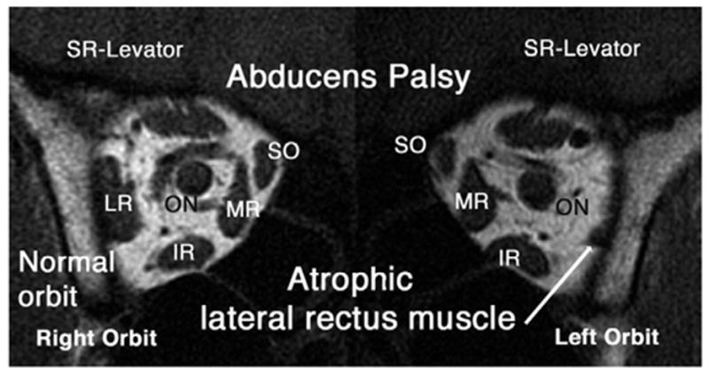
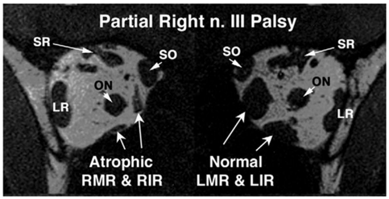
Methods: Two men (ages 31 and 47 years) and 3 women (ages 40-49 years) with CPEO and symptom durations of 8 months to 28 years underwent high-resolution (2-mm slice thickness, 312 micron pixels), surface coil, T1-weighted orbital MRI in coronal planes. Images were analyzed quantitatively to determine extraocular muscle (EOM) sizes and were compared with 10 age- and gender-matched normal volunteers, one subject with myasthenia gravis, and with 30 subjects having EOM paralysis caused by oculomotor, trochlear,0 and abducens neuropathies.
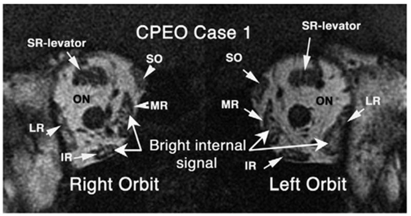
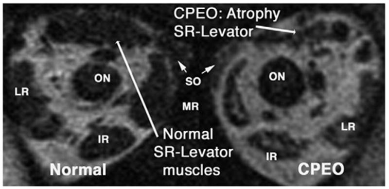
Results: EOM function was clinically diminished in CPEO, most markedly for the superior rectus (SR) and levator muscles. All EOMs in CPEO exhibited unusual qualitative T1 MRI signal abnormalities. Unlike the profound EOM atrophy typical of neurogenic paralysis, anterior volumes of medial rectus, lateral rectus, and inferior rectus muscles in CPEO were not smaller than normal (p>0.003). Anterior volumes of the SR muscle-levator complex and superior oblique were significantly reduced (p<0.003). Denervated EOMs exhibited statistically significant volume reduction when compared with normal and CPEO groups. Volume of the SR muscle-levator complex was the same in subjects with CPEO and oculomotor palsies.
Conclusions: CPEO is associated with minimal EOM volume reduction despite clinically severe weakness. This combination of findings may be specific for CPEO and could resolve the diagnostic dilemma in difficult cases.
Figures
Similar articles
-
MR of extraocular muscles in chronic progressive external ophthalmoplegia.AJNR Am J Neuroradiol. 1998 Jan;19(1):95-9. AJNR Am J Neuroradiol. 1998. PMID: 9432164 Free PMC article.
-
Extra-ocular muscle MRI in genetically-defined mitochondrial disease.Eur Radiol. 2016 Jan;26(1):130-7. doi: 10.1007/s00330-015-3801-5. Epub 2015 May 21. Eur Radiol. 2016. PMID: 25994195 Free PMC article.
-
Eye Muscle MRI in Myasthenia Gravis and Other Neuromuscular Disorders.J Neuromuscul Dis. 2023;10(5):869-883. doi: 10.3233/JND-230023. J Neuromuscul Dis. 2023. PMID: 37182896 Free PMC article.
-
Extraocular mitochondrial myopathies and their differential diagnoses.Strabismus. 2006 Jun;14(2):107-13. doi: 10.1080/09273970600701218. Strabismus. 2006. PMID: 16760117 Review.
-
False positive acetylcholine receptor antibodies in a case of unilateral chronic progressive external ophthalmoplegia: case report and review of literature.Orbit. 2018 Oct;37(5):385-388. doi: 10.1080/01676830.2017.1423350. Epub 2018 Jan 15. Orbit. 2018. PMID: 29333908 Review.
Cited by
-
Radiological Characteristics of Extraocular Muscles in Myasthenia Gravis Patients with Ocular Manifestations: A Case-Control Study.Clin Ophthalmol. 2021 Jun 1;15:2279-2285. doi: 10.2147/OPTH.S280508. eCollection 2021. Clin Ophthalmol. 2021. PMID: 34103891 Free PMC article.
-
MRI evidence of extraocular muscle atrophy and fatty replacement in myasthenia gravis.Neuroradiology. 2021 Sep;63(9):1531-1538. doi: 10.1007/s00234-021-02753-4. Epub 2021 Jul 7. Neuroradiology. 2021. PMID: 34232334
-
Selected lid problems in neurologic practice.Curr Neurol Neurosci Rep. 2009 Sep;9(5):390-5. doi: 10.1007/s11910-009-0057-y. Curr Neurol Neurosci Rep. 2009. PMID: 19664369 Review.
-
A systematic review on volumetric analysis in orbital MRI.Neuroradiology. 2025 Sep 6. doi: 10.1007/s00234-025-03734-7. Online ahead of print. Neuroradiology. 2025. PMID: 40913736 Review.
-
Progressive External Ophthalmoplegia.Curr Neurol Neurosci Rep. 2016 Jun;16(6):53. doi: 10.1007/s11910-016-0652-7. Curr Neurol Neurosci Rep. 2016. PMID: 27072953 Review.
References
-
- Von Graefe A. Verhandlungen arztlicher Gesselschaften. Berlin Klin Wochenschr. 1868;5:125.
-
- Miller NR, editor. Walsh and Hoyt’s clinical neuro-ophthalmology. 4. Vol. 2. Baltimore (MD): Williams and Wilkins; 1985. pp. 811–23.
-
- Rowland LP. Progressive external ophthalmoplegia and ocular myopathies. In: Rowland LP, Di Mauro S, editors. Handbook of clinical neurology, revised series. Vol. 18. Amsterdam: Elsevier; 1992. pp. 287–329.
-
- Biousse V, Newman NJ. Neuro-ophthalmology of mitochondrial diseases. Semin Neurol. 2001;21:275–291. - PubMed
-
- Biousse V, Newman NJ. Neuro-ophthalmology of mitochondrial diseases. Curr Opin Neurol. 2003;16:35–43. - PubMed
Publication types
MeSH terms
Grants and funding
LinkOut - more resources
Full Text Sources
Medical
Research Materials
