. 2007 Feb 1;302(1):256-66.
doi: 10.1016/j.ydbio.2006.09.048. Epub 2006 Oct 4.

Periostin promotes atrioventricular mesenchyme matrix invasion and remodeling mediated by integrin signaling through Rho/PI 3-kinase

Affiliations

Periostin promotes atrioventricular mesenchyme matrix invasion and remodeling mediated by integrin signaling through Rho/PI 3-kinase

Jonathan T Butcher et al. Dev Biol. .

Abstract

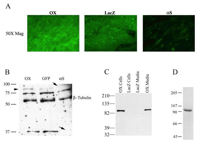
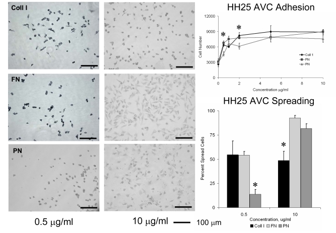
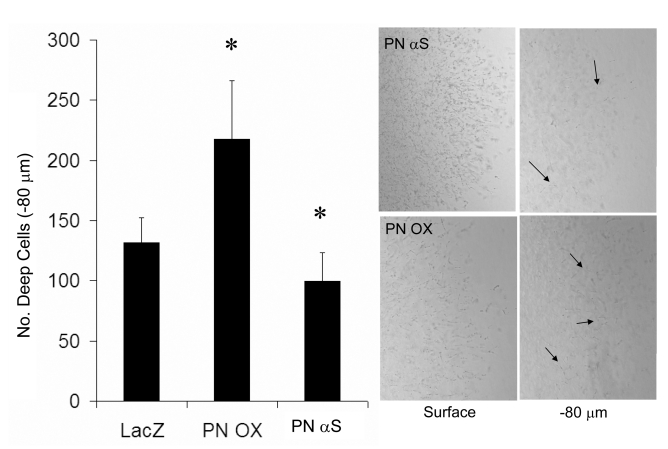
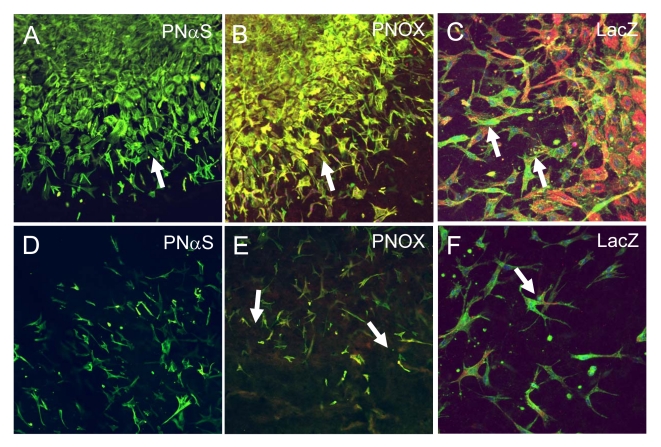
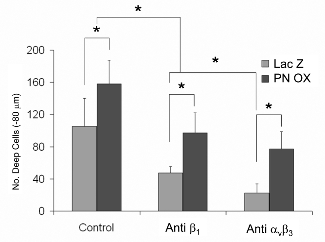
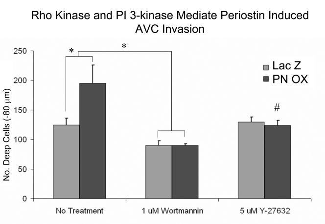
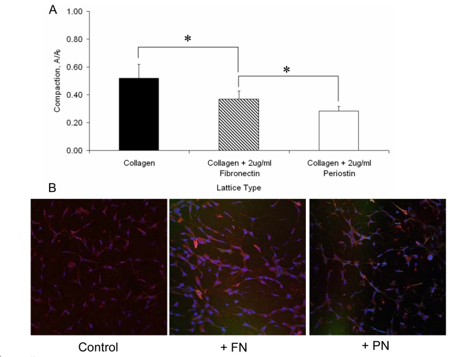
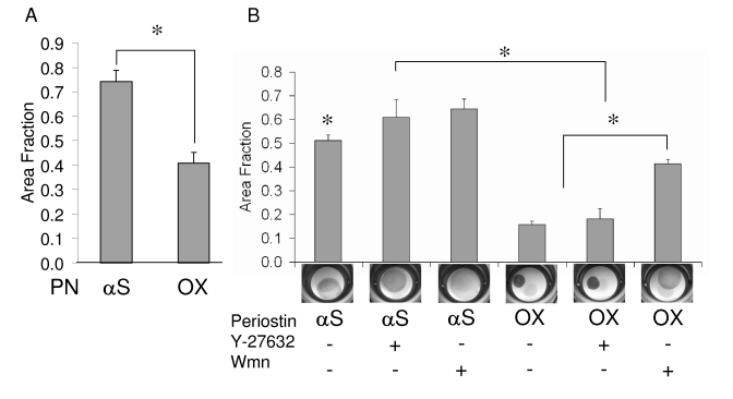
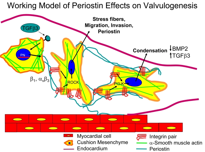
Recent evidence suggests that extracellular matrix components may play a signaling role in embryonic valve development. We have previously identified the spatiotemporal expression patterns of periostin in developing valves, but its function during this process is largely unknown. To evaluate the functional role periostin plays during valvulogenesis, two separate three-dimensional culture assay systems, which model chick atrioventricular cushion development, were employed. These assays demonstrated that cushion mesenchymal cells adhered and spread on purified periostin in a dose-responsive manner, similar to collagen I and fibronectin via alpha(v)beta(3) and beta(1) integrin pairs. Periostin overexpression resulted in enhanced mesenchyme invasion through 3D collagen gels and increased matrix compaction. This invasion was dependent on alpha(v)beta(3) more than beta(1) integrin signaling, and was mediated differentially by Rho kinase and PI 3-kinase. Both matrix invasion and compaction were associated with a colocalization of periostin and beta(1) integrin expression to migratory cell phenotype in both surface and deep cells. The Rho/PI 3-kinase pathway also differentially mediated matrix compaction. Both Rho and PI 3-kinase were involved in normal cushion mesenchyme matrix compaction, but only PI 3-kinase was required for the enhanced matrix compaction due to periostin. Taken together, these results highlight periostin as a mediator of matrix remodeling by cushion mesenchyme towards a mature valve structure.


Figures

Fig. 1
Fig. 1
Validation of periostin virus expression and protein purification. A: Immunohistochemical validation of periostin overexpression and knockdown in HH26 outflow tract cells by sense and antisense adenovirus as compared to LacZ control. A significant increase in periostin positive cells is evident in the PNOX cells whereas little appreciable staining is seen in the αS B: Western blot of infected HEK293 cells with the periostin overexpressing adenovirus. Expression of the 90kDa adenovirally produced periostin protein is clearly demonstrated in the HEK293 supernatant. Non-infected control shows antibody specificity. C. Functional validation of the periostin adenoviruses in HH25 AV cushion tissue. An overexpression (2.2 net increase) of the 90kDa adenovrially expressed variant of periostin (OX) is seen when compared to the endogenous levels (GFP). Infection with the αS periostin adenovirus resulted in a complete absence of the 90kDa and the 37 kDa variants. No change was observed in a 75 kDa periostin variant. β-tubulin was used as a normalization control. For both blots (B & C) the α-mouse periostin antibody was utilized. D: Purification of full length periostin from HEK293 cells determined by Western blot. Two μg of periostin purified as described in the Materials and Methods was resolved by SDS-PAGE and detected by Coomassie blue staining. Only one band was present at the 90 kDa, demonstrating the purity of the periostin preparation.
Fig. 2
Fig. 2
Adhesion and spreading of HH25 Atrioventricular cushion mesenchyme to different doses of extracellular matrix proteins type I collagen (Coll I) fibronectin (FN) and periostin (PN). * denotes significance at P<0.05.
Fig. 3
Fig. 3
Adhesion to periostin (but not fibronectin) is dependent on αvβ3 and β1 integrins. 30,000 cushion mesenchyme cells were allowed to adhere for 1 hour in the presence of blocking antibodies used at 5 μg/ml concentration. * denotes significant difference between matrix proteins (P<0.05), while # denotes significant between treatments (P<0.05).
Fig. 4
Fig. 4
Periostin overexpression enhances cushion mesenchyme invasion (arrows) over LacZ controls while antisense reduces invasion. * denotes significance P<0.05.
Fig. 5
Fig. 5
Immunohistochemistry of surface (0 μm, panels A-C) and deep (−80 μm, panels D-F) sections of invading HH25 AV cushion mesenchyme. PNαS infected cultures exhibited almost no detection of periostin (red) in either surface (A) or deep cells (D). PNOX infected cultures coexpressed periostin (red) and a-smooth muscle actin (green) in migratory/invasive morphology in both surface (B) and deep cells (E). β1 integrin (green) expression was also co-localized with a-smooth muscle actin (red) in migratory/invasive cells at both surface (C) and deep levels (F). All images are at 200x magnification.
Fig. 6
Fig. 6
Cushion mesenchyme invasion into collagen I gels is dependent on integrins. Blocking antibodies to αvβ3 or β1 integrins (5 μg/ml) were added to culture medium and resulted in reduced invasion from both LacZ control and periostin overexpressing cultures. * denotes significance at P<0.05.
Fig. 7
Fig. 7
Cushion mesenchyme invasion into collagen I gels is dependent on Rho/PI 3-kinase. PI 3-kinase inhibitor (Wortmannin) resulted in reduced numbers of invaded cells over controls, with a greater reduction of invasion in PNOX cultures. Rho kinase was not involved in control cell invasion, but inhibition of Rho kinase with Y-27632 in PNOX cultures results in a reduction of invaded cells back to control levels. * denotes significance at P<0.05, # denotes significantly different from no treatment control.
Fig. 8
Fig. 8
Condensation of collagen I matrix by HH25 AV cushion mesenchyme. (A) Supplementation with purified fibronectin (FN, 2 μg/ml) or periostin (PN, 2μg/ml) resulted in enhanced condensation, with periostin supplemented constructs compacting the most. (B) Enhanced matrix condensation is associated with increased expression of α-smooth muscle actin. FN and PN supplemented collagen I gels had significantly greater expression compared to collagen I only controls. * denotes significance at P<0.05.
Fig. 9
Fig. 9
Effects of Rho/PI 3-kinase inhibition on matrix condensation by periostin overexpressing cushion mesenchyme. A: Increased matrix compaction after 7 days by periostin overexpressing constructs compared to antisense periostin. B: Matrix compaction after an additional three days incubation with Rho kinase inhibitor Ỹ-27632 (5 μM) or PI 3-kinase inhibitor Wortmannin (Wmn, 1 μM). “+” denotes addition of the inhibitor, “-“ without inhibitor. αS = antisense, OX = overexpression. * denotes significance P<0.05.
Fig. 10
Fig. 10
Schematic detailing the proposed roles of periostin in mediating post-EMT cushion remodeling. After EMT (approximately HH20 in chick) a signaling ligand (our preliminary evidence suggests it is TGFβ3) signals mesenchymal cells to synthesize and secrete periostin. Mesenchyme then adhere to periostin via β1 and αvβ3 integrin pairs and further invade and migrate within the collagenous cushion matrix via Rho kinase signaling, stress fiber formation, and filipodia extension. By HH25, cushion tissue is fully mesenchymalized and the resident cells begin to condense the matrix, this process enhanced by periostin signaling via PI-3 kinase. This condensation contributes to thinning the cushions into fibrous leaflets and delaminating the tisuue from the myocardial wall by an as yet unknown mechanism, but possibly related to the upregulation of TGFβ3 and downregulation of BMP2 in chick.

References

    1. Baldwin HS, Lloyd TR, Solursh M. Hyaluronate degradation affects ventricular function of the early postlooped embryonic rat heart in situ. Circ Res. 1994;74:244–52. - PubMed
    1. Banyard J, Anand-Apte B, Symons M, Zetter BR. Motility and invasion are differentially modulated by Rho family GTPases. Oncogene. 2000;19:580–91. - PubMed
    1. Borck A, Massey E, Loftis MJ, Carver W. Exposure of cardiac fibroblasts to the herbicide nitrofen causes altered interactions with the extracellular matrix. Cell Biol Toxicol. 2004;20:15–24. - PubMed
    1. Bornstein P, Sage EH. Matricellular proteins: extracellular modulators of cell function. Curr Opin Cell Biol. 2002;14:608–16. - PubMed
    1. Butcher JT, Penrod AM, Garcia AJ, Nerem RM. Unique morphology and focal adhesion development of valvular endothelial cells in static and fluid flow environments. Arterioscler Thromb Vasc Biol. 2004;24:1429–34. - PubMed

Publication types

MeSH terms