. 2007 Mar;204(1):77-87.
doi: 10.1016/j.expneurol.2006.09.018. Epub 2006 Oct 27.

Long-lasting impairment in hippocampal neurogenesis associated with amyloid deposition in a knock-in mouse model of familial Alzheimer's disease

Affiliations

Long-lasting impairment in hippocampal neurogenesis associated with amyloid deposition in a knock-in mouse model of familial Alzheimer's disease

Chen Zhang et al. Exp Neurol. 2007 Mar.

Abstract

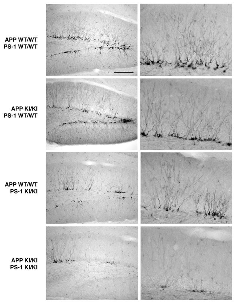
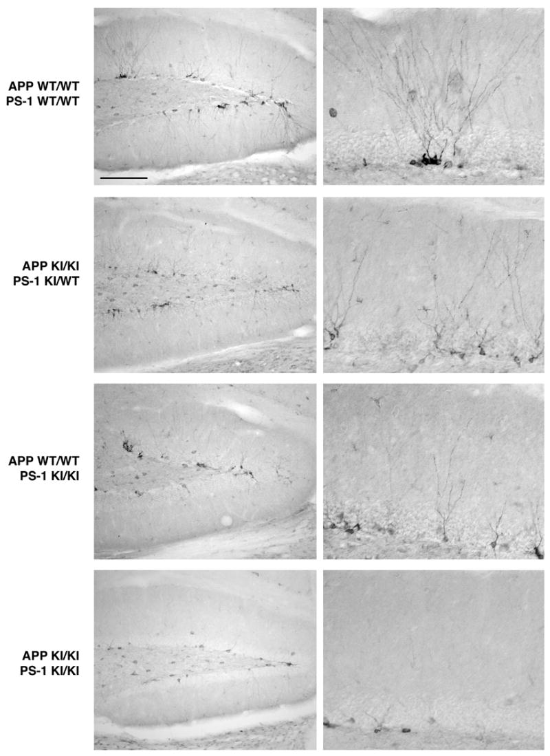
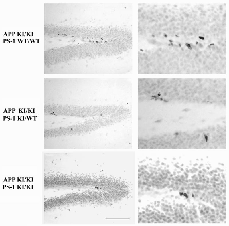
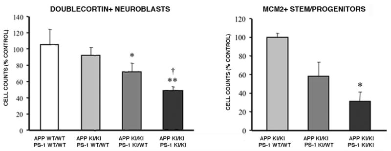
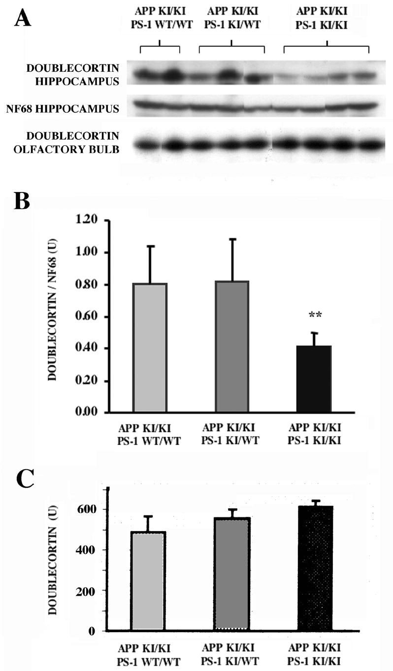
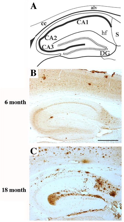
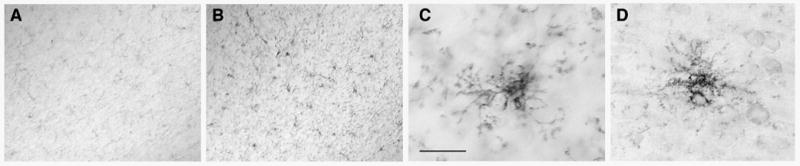
Neurogenesis in the adult hippocampus has been implicated in regulating long-term memory and mood, but its integrity in Alzheimer's disease (AD) is uncertain. Studies of neurogenesis in transgenic mouse models of familial AD are complicated by ectopic overexpression restricted to terminally differentiated neurons, while AD cases have been studied only at the pre-senile or end-stage of disease. To investigate further the fidelity of adult neurogenesis, we examined mice carrying targeted mutations in amyloid precursor protein (APP), presenilin-1 (PS-1), or both APP and PS-1, in which FAD-causing mutations have been inserted into their endogenous genes. The latter "double knock-in" mice developed aging- and region-dependent amyloid deposition starting around 6 months, and by 9 months exhibited microglial activation associated with the amyloid. In the 9-month-old dentate gyrus, the double knock-in mutations reduced the numbers of MCM2-positive neural stem and progenitor cells by 3-fold and doublecortin-positive neuroblasts by 2-fold. The reduction in dentate neuroblasts persisted at 18 months of age. The impairment in neurogenesis was confirmed by quantitative Western blot analysis of doublecortin content and was restricted to the hippocampal but not the olfactory bulb neurogenic system. In contrast, neither mutant PS-1 nor APP alone led to amyloid deposition or significant alterations in the two markers. These results demonstrate long-lasting and selective impairment in adult hippocampal neurogenesis in a knock-in mutant mouse model of FAD and suggest a novel mechanism by which amyloid and its attendant microglia-mediated neuroinflammation could contribute to the cognitive and behavioral abnormalities of AD.


Figures

Figure 1
Figure 1
Developing neurons in adult hippocampus: effects of FAD-linked knock-in mutations in APP, PS-1, and both genes. Sagittal sections from 8–9 month old mice of the indicated genotypes were immunostained for the neuroblast marker doublecortin. Low (left panels) and high (right panels) magnification views of the dentate gyrus at a mid-mediolateral level. Note the abundance of immature neurons in the subgranular zone adjacent to the granule cell layer of both the suprapyramidal and infrapyramidal blades of the dentate gyrus. The homozygous APP Swedish knock-in mutation had no effect on immunopositive neuroblasts, whereas the homozygous PS-1 P264L mutation led to a small decrease in doublecortin-positive neuroblast number. In contrast to the effects of the mutations alone, the combined homozygous mutations in both APP and PS-1 caused a marked reduction in the number of developing neurons. Scale bar = 200 μm (left panels), 50 μm (right panels).
Figure 2
Figure 2
Defective neurogenesis in the APP/PS-1 double knock-in mutant mouse hippocampus persists during aging. Sagittal sections from 18–20 month old mice of the indicated genotypes immunostained for doublecortin. With aging, there is a reduction in the number of developing neurons in the subgranular zone when compared with the 8–9 month age. As seen for the earlier age, the APP knock-in mutation does not affect neuroblast number and the PS-1 knock-in mutation causes only a small decrease. On the other hand, the APP/PS-1 double knock-in mutation led to a large reduction in the number of developing neurons. Scale bar = 200 μm (left panels), 50 μm (right panels).
Figure 3
Figure 3
The PS-1 knock-in mutation reduces the number of neural stem and progenitor cells in the hippocampus when combined with the APP knock-in mutation. Sagittal sections from 8–9 month old mice of the indicated genotypes immunostained for the marker of cycling cells MCM2. Cells intensely stained for this marker are found along the dentate subgranular zone. For mice homozygous for the APP Swedish knock-in mutation, the addition of one mutant PS-1 allele caused a small decrease in immunopositive neural stem and progenitor cells. Two mutant PS-1 alleles led to a further decline in these cell populations. Scale bar = 100 μm (A,B), 40 μm (C,D).
Figure 4
Figure 4
Quantitative analysis of neuroblasts and neural stem/progenitors as functions of APP and PS-1 genotype. The numbers of doublecortin-positive immature neurons and MCM2-positive neural stem and progenitors were measured for the entire dorsal extent of the hippocampal dentate gyrus, as described in Materials and Methods. When combined with the homozygous APP Swedish knock-in mutation, one PS-1 P264L mutant allele caused a small decline in both types of cell populations. Significant decreases in the neuroblast and neural stem/progenitor populations of greater magnitude were observed for the APP/PS-1 double homozygous knock-in mutation. (A) *p<0.05, **p<0.01 versus wild type mice, †p<0.01 versus APP KI/KI + PS-1 WT/WT mice; (B) *p<0.05 versus APP KI/KI + PS-1 WT/WT mice, ANOVA post hoc comparison.
Figure 5
Figure 5
Western blot analysis of doublecortin content of hippocampus and olfactory bulb as a function of APP and PS-1 genotype. The regions were dissected from 8–9 month old mice of the indicated APP and PS-1 genotypes, and the extracts probed for the ~42 kDa doublecortin polypeptide as described in Materials and Methods. In addition, the hippocampal samples were reprobed for neurofilament L (NF68). (A) Western blots illustrating that the APP/PS-1 double knock-in mutation decreased hippocampal doublecortin content when compared with APP knock-in mutant mice bearing either zero or one mutant PS-1 allele. (B,C) Quantitative analysis of doublecortin content for hippocampus (B) and olfactory bulb (C). Hippocampal doublecortin levels are represented as the ratio of the densities of the doublecortin and neurofilament L polypeptides. Essentially identical results were obtained from analysis of doublecortin alone. The decrease in doublecortin levels in the hippocampus of the APP/PS-1 double knock-in mutant mouse is significant (p<0.05, t-test; p=.06, ANOVA). In marked contrast, there was no appreciable difference as a function of PS-1 gneotype in doublecortin content of the olfactory bulb.
Figure 6
Figure 6
Amyloid deposition is extensive in the dentate gyrus of the APP/PS-1 double knock-in mutant mouse. Sagittal sections from either 6 (B) or 18 (C) month old APP/PS-1 double knock-in mutant mice were immunostained for amyloid Aβ. (A) Schematic diagram of mouse dorsal hippocampus in the sagittal plane at mid-mediolateral level. Amyloid deposits became detectable in the dentate gyrus starting at 6 months of age. By 18 months, amyloid plaque formation was extensive especially in the outer molecular layer of the dentate gyrus. Scale bar = 500 μm.
Figure 7
Figure 7
Microglial activation in association with amyloid deposition in the APP/PS-1 double knock-in mutant mouse. Sagittal sections of 18 month old mouse dentate gyrus immunostained for the activated microglial markers F4/80 (A–C) and CD45 (D). (A) APP KI/KI + PS-1 WT/WT. (B–D) APP KI/KI + PS-1 KI/KI. The numbers of cells immunostained for the markers of activated microglia were markedly higher for the double knock-in mutant mouse hippocampus. At high magnification (C,D), immunopositive cells had the small cell bodies and thin, highly ramified processes characteristic for microglia. Scale bar = 200 μm (A,B), 25 μm (C,D).

References

    1. Aimone JB, Wiles J, Gage FH. Potential role for adult neurogenesis in the encoding of time in new memories. Nat Neurosci. 2006;9:723–727. - PubMed
    1. Ashe KH. Learning and memory in transgenic mice modeling Alzheimer's disease. Learn Mem. 2001;8:301–308. - PubMed
    1. Bornemann KD, Wiederhold KH, Pauli C, Ermini F, Stalder M, Schnell L, Sommer B, Jucker M, Staufenbiel M. Abeta-induced inflammatory processes in microglia cells of APP23 transgenic mice. Am J Pathol. 2001;158:63–73. - PMC - PubMed
    1. Braak H, de Vos RA, Jansen EN, Bratzke H, Braak E. Neuropathological hallmarks of Alzheimer's and Parkinson's diseases. Prog Brain Res. 1998;117:267–285. - PubMed
    1. Cameron HA, McKay RD. Restoring production of hippocampal neurons in old age. Nat Neurosci. 1999;2:894–897. - PubMed

Publication types

MeSH terms