. 2007 Jan 12;365(2):535-50.
doi: 10.1016/j.jmb.2006.10.011. Epub 2006 Oct 7.

Determining the critical nucleus and mechanism of fibril elongation of the Alzheimer's Abeta(1-40) peptide

Affiliations

Determining the critical nucleus and mechanism of fibril elongation of the Alzheimer's Abeta(1-40) peptide

Nicolas Lux Fawzi et al. J Mol Biol. .

Abstract

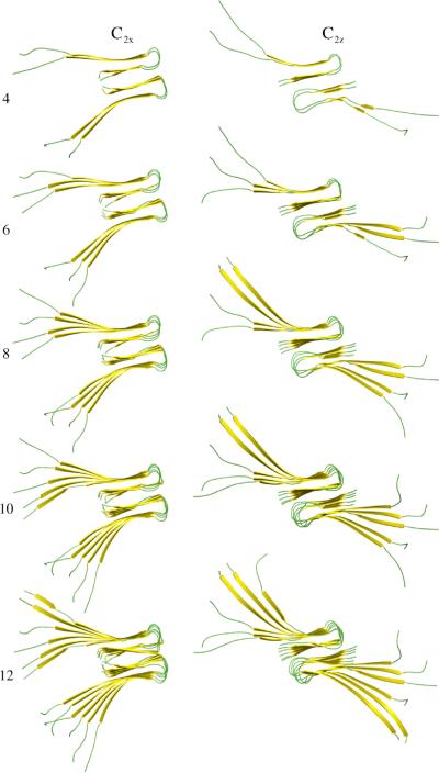
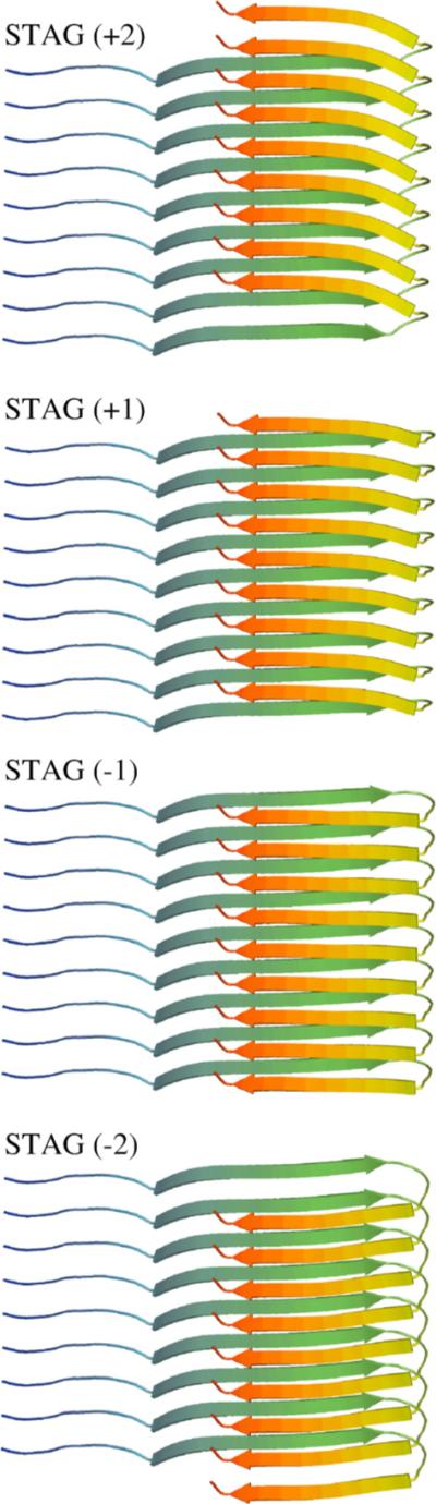
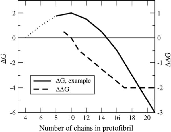
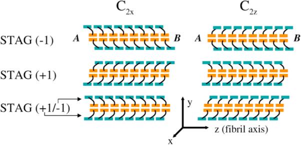
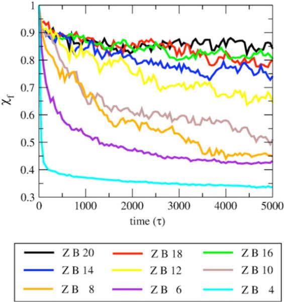
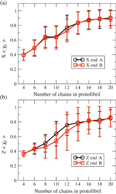
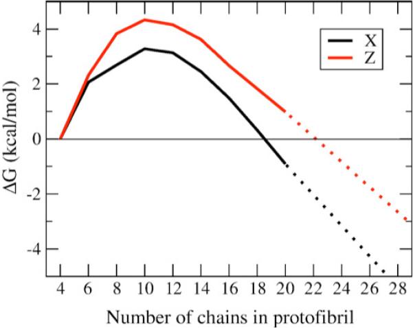
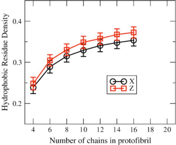
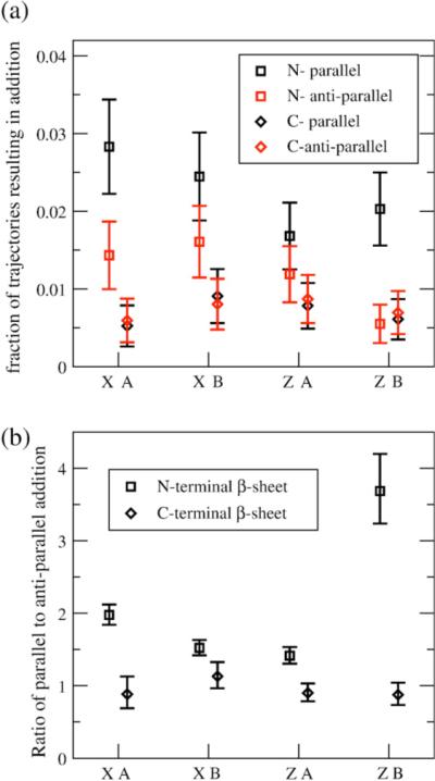
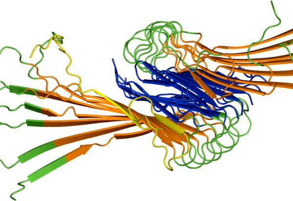
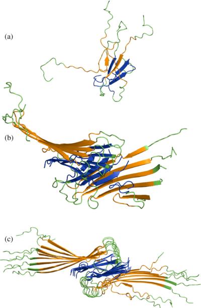
We use a coarse-grained protein model to characterize the critical nucleus, structural stability, and fibril elongation propensity of Abeta(1-40) oligomers for the C(2x) and C(2z) quaternary forms proposed by solid-state NMR. By estimating equilibrium populations of structurally stable and unstable protofibrils, we determine the shift in the dominant population from free monomer to ordered fibril at a critical nucleus of ten chains for the C(2x) and C(2z) forms. We find that a minimum assembly of 16 monomer chains is necessary to mimic a mature fibril, and show that its structural stability correlates with a plateau in the hydrophobic residue density and a decrease in the likelihood of losing hydrophobic interactions by rotating the fibril subunits. While Abeta(1-40) protofibrils show similar structural stability for both C(2x) and C(2z) quaternary structures, we find that the fibril elongation propensity is greater for the C(2z) form relative to the C(2x) form. We attribute the increased propensity for elongation of the C(2z) form as being due to a stagger in the interdigitation of the N-terminal and C-terminal beta-strands, resulting in structural asymmetry in the presented fibril ends that decreases the amount of incorrect addition to the N terminus on one end. We show that because different combinations of stagger and quaternary structure affect the structural symmetry of the fibril end, we propose that differences in quaternary structures will affect directional growth patterns and possibly different morphologies in the mature fiber.


Figures

Figure 1
Figure 1
Examples of starting structures for C2x and C2z symmetry forms. Protofibril seeds composed of four, six, eight, ten, and 12 (14−20 not shown) monomers were simulated for protofibril stability.
Figure 2
Figure 2
Interdigitation of the N and C-terminal β-strands to form side-chain contacts between different monomer chains introduces a stagger in the strand alignments. Side-chain contacts (from top to bottom) between the N termini of monomer i with the C termini of monomers i + 1 and i + 2 (STAG (+2)), between the N termini of i with the C termini of i and i + 1 (STAG (+1)), between the C termini of monomer i with the N termini of monomer i and i + 1 (STAG (−1)), or between the C termini of i with the N termini of i + 1 and i + 2 (STAG (−2)). Our model relaxes naturally to the STAG (−1) definition.
Figure 3
Figure 3
Free energy profile for the nucleation-polymerization reactions. Typical free energy (ΔG) profile and slope of ΔG (ΔΔG) versus the number of chains in the protofibril for fibril formation by a nucleation-dependent polymerization mechanism. At high numbers of chains, the protofibril is stable and free energetically favorable, and the free energy benefit to adding chains is constant, as seen in a constant slope of ΔG. Since the slope of ΔG is constant in this regime, the free energy benefit to adding a chain or free energy cost for removing a chain is the same as in an infinite fibril. As the number of chains decreases, the free energy change for removing chains decreases, indicating that the fibril is approaching the number of chains in the critical nucleus. At the critical nucleus, the least free energetically favorable species, the slope of ΔG is zero. (Typical ΔG data adapted from Ferrone23).
Figure 4
Figure 4
Effect of internal stagger on terminating ends of fibril. A schematic of 16 chain C2x and C2z fibrils are shown for internal staggers STAG (−1), STAG (+1) and mixed STAG (+1/−1) with the N-terminal region colored teal and the C-terminal region colored orange. STAG (−1) C2x has superimposable, symmetric ends. End A can be approximately superimposed on end B by a simple rotation of 180° about the x-axis (hence C2x). STAG (−1) C2z has distinct, asymmetric ends. End A exposes the C-terminal β-strands, and end B exposes the N-terminal β-strands. Ends A and B of the C2z fibril cannot be superimposed on end A by any rotation. C2x STAG (+1), like C2x STAG (−1), has superimposable, symmetric ends. C2z STAG (+1), like C2z STAG (−1) above it, has distinct, asymmetric ends. C2x STAG (−1/+1) has the top peptide STAG (+1) and bottom peptide STAG (−1). Mixing staggers in C2x de-symmetrizes the C2x ends. Mixing staggers in C2z symmetrizes the C2z ends, so that each end has one subunit with an exposed N-terminal β-strand, and the other with an exposed C-terminal β-strand, unlike the two asymmetric ends in “pure” C2z STAG (−1) or C2z STAG (+1) models.
Figure 5
Figure 5
Time-course for protofibril stability measured by χf. The metric χf measures the pair distances between the residues on both sides of the fibril, and is more sensitive to rotation of one subunit with respect to the other, and thus measures fibril disorder of the quaternary structure. The time-course data averaged over all trajectories of C2z fibrils for lengths four to 20 chains for fibril end B.
Figure 6
Figure 6
Protofibril stability measured by <χf> versus number of chains; <Χf> is an average measure of the fibril order of the edge chains for stable quaternary structure for (a) C2x form and (b) C2z for the two ends of the protofibril: end A (black) and end B (red). Note the difference between the two distinguishable ends of the C2z oligomers due to stagger effects. The error bars represent standard deviation.
Figure 7
Figure 7
Free energy profile for free monomer and protofibril equilibrium. The free energy versus number of ordered chains in an oligomer is plotted for C2x (X, black) and C2z forms. The free energy shows a clear maximum at ten chains for C2z and ten to 12 chains for C2x, indicating the region of the critical nucleus. A constant, negative slope at ∼16 chains and above is indicative of reaching a stable fibril regime.
Figure 8
Figure 8
Hydrophobic residue density versus number of chains. Hydrophobic density (number of hydrophobic residues per unit volume) versus number of chains for the C2x and C2z forms after initial equilibration. The error bars represent standard deviation for the 24 structures created from the 40 chain equilibration runs. The hydrophobic density for C2z is higher than C2x for all oligomer sizes.
Figure 9
Figure 9
Monomer additions to protofibrils for C2x and C2z fibrils. (a) Fraction of trajectories resulting in partial parallel (black) and antiparallel (red) additions to the N-terminal (□) and C-terminal (◇) β-sheets. The error bars represent standard deviation approximated from distributions with binary outcomes. (b) Ratio of partial parallel to antiparallel additions to the N-terminal (□) and C-terminal (◇) β-sheets. The error bars are the 95% confidence interval for “relative risk” measure comparing binary outcomes.
Figure 10
Figure 10
Example addition to fibril seed by free peptide. A peptide (yellow) with a random initial configuration without contacts with the seed is shown with partial in-register parallel addition to both N-terminal and C-terminal β-sheets of the fibril seed.
Figure 11
Figure 11
Comparing structural stability of example structures of varying length of oligomer. Representative oligomer structures after 5000τ constant temperature simulations depicting greater structural stability as number of chains increases. (a) Four-chain simulation shows a complete loss of fibril structure. (b) Ten-chain simulation shows that, although a significant fraction of intermolecular β-sheet is retained, the fibril subunits rotate with respect to one another, leading to disorder and loss of contacts in the edge chains. (c) The 16-chain simulations show retention of fibril order, and a clear fibril axis.

References

    1. Dobson CM. Protein folding and misfolding. Nature. 2003;426:884–890. - PubMed
    1. Dobson CM. Principles of protein folding, misfolding and aggregation. Semin. Cell Dev. Biol. 2004;15:3–16. - PubMed
    1. Bucciantini M, Giannoni E, Chiti F, Baroni F, Formigli L, Zurdo J, et al. Inherent toxicity of aggregates implies a common mechanism for protein misfolding diseases. Nature. 2002;416:507–511. - PubMed
    1. Stefani M, Dobson CM. Protein aggregation and aggregate toxicity: new insights into protein folding, misfolding diseases and biological evolution. J. Mol. Med. 2003;81:678–699. - PubMed
    1. Cohen FE, Kelly JW. Therapeutic approaches to protein-misfolding diseases. Nature. 2003;426:905–909. - PubMed

Publication types

LinkOut - more resources