. 2006 Nov;169(5):1633-42.
doi: 10.2353/ajpath.2006.060501.

Insulin-like growth factor-binding protein-5 induces pulmonary fibrosis and triggers mononuclear cellular infiltration

Affiliations

Insulin-like growth factor-binding protein-5 induces pulmonary fibrosis and triggers mononuclear cellular infiltration

Hidekata Yasuoka et al. Am J Pathol. 2006 Nov.

Abstract

We have recently shown that insulin-like growth factor-binding protein (IGFBP)-5 is overexpressed in idiopathic pulmonary fibrosis lung tissues and increases collagen and fibronectin deposition. Here, we further examined the effect of IGFBP-5 in vivo by intratracheal administration of replication-deficient adenovirus expressing human IGFBP-5 (Ad5), IGFBP-3 (Ad3), or no cDNA (cAd) to wild-type mice. Increased cellular infiltration and extracellular matrix deposition were observed in mice after Ad5 administration compared with Ad3 and cAd. Mononuclear cell infiltration consisted predominantly of T lymphocytes at day 8. By day 14, the number of infiltrating T cells decreased, whereas that of B cells and monocytes/macrophages increased. IGFBP-5 also induced migration of peripheral blood mononuclear cells in vitro, suggesting that in vivo mononuclear cell infiltration may be the direct result of IGFBP-5 expression. alpha-Smooth muscle actin and Mucin-1 co-localized in cells of mice treated with Ad5, suggesting that IGFBP-5 induced epithelial-mesenchymal transition. In addition, exogenous IGFBP-5 induced alpha-smooth muscle actin expression in primary fibroblasts and epithelial-mesenchymal transition of pulmonary epithelial cells in vitro. In conclusion, our results suggest that overexpression of IGFBP-5 in mouse lung results in fibroblast activation, increased extracellular matrix deposition, and myofibroblastic changes. Thus, the IGFBP-5-induced fibrotic phenotype in vivo may represent a novel model to better understand the pathogenesis of fibrosis.


Figures
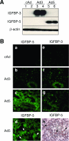
Figure 1
Figure 1
A: IGFBP-3 and IGFBP-5 secretion by cultured primary mouse lung fibroblasts infected with control adenovirus (cAd, lanes 1 and 2), IGFBP-3-expressing (Ad3, lanes 3 and 4), and IGFBP-5-expressing adenovirus (Ad5, lanes 5 and 6). B: IGFBP-3 and IGFBP-5 expression in mouse lung. Control adenovirus (cAd) or adenovirus expressing human IGFBP-3 and IGFBP-5 (Ad3 and Ad5, respectively) was administered intratracheally. Mice were sacrificed and lung tissues harvested 14 days after injection. Expression of IGFBP-5 (a–d) and IGFBP-3 (e–g) was detected using immunofluorescence and that of IGFBP-5 was also detected by immunohistochemistry (h). Original magnifications, ×400.
Figure 2
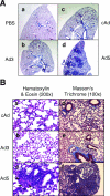
Figure 2
In vivo effects of IGFBP-3 and IGFBP-5 overexpression in mouse lung. A: Increased collagen deposition in IGFBP-5-expressing lungs. Phosphate-buffered saline (PBS) (a), 109 PFU control adenovirus (cAd) (c), or IGFBP-3- and IGFBP-5-expressing adenovirus [Ad3 (b) and Ad5 (d), respectively] were administered intratracheally to mice. Mice were sacrificed 2 weeks later, and lung tissue sections were stained with Masson’s Trichrome. B: Mononuclear cell infiltration and ECM deposition in IGFBP-5-expressing lungs. Lung sections from cAd-, Ad3-, and Ad5-treated mice were stained with H&E (a–c) or Masson Trichrome staining (d–f). Original magnifications: ×20 (A); ×200 (Ba–Bc); ×100 (Bd–Bf).
Figure 3
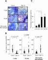
Figure 3
Characterization of infiltrating immune cells. A: Immunohistochemistry for the detection of T lymphocytes, B lymphocytes, and monocytes/macrophages. Mice were infected with 109 PFU control-, IGFBP-3-, or IGFBP-5-expressing adenovirus (cAd, Ad3, and Ad5, respectively), and lung samples were analyzed at 8 days and 14 days after injection. Solid arrows point to representative positive cells. B: Number of T cells, B cells, and monocytes/macrophages in mouse lungs. Two representative samples were randomly selected from each group of mice and the number of each cell subset was counted in 10 random fields/sample at ×400 magnification. Statistical analysis was done using Student’s t-test. Each bar denotes the mean of each group. C: PBMC migration assay. Purified human PBMCs were used in a migration assay with vehicle control, recombinant IGFBP-3 (BP3), recombinant human IGFBP-5 (BP5), or RANTES as a positive control. Statistical analysis was done using the Mann-Whitney U-test. *P < 0.007. Original magnifications, ×400.
Figure 4
Figure 4
A: Collagen production and deposition in lung tissues. The amount of collagen was measured in lungs injected with 109 PFU control adenovirus-, IGFBP-3-, or IGFBP-5-expressing adenoviruses (cAd, Ad3, and Ad5, respectively) using a hydroxyproline assay. The bar in each group denotes mean collagen amount. Statistical analysis was done using the Mann-Whitney U-test. *P < 0.03. B: Expression of fibronectin and α-SMA in mouse lungs 14 days after injection. Expression of fibronectin and α-SMA was examined in mouse lung samples infected with 109 PFU control adenovirus (cAd), IGFBP-3- or IGFBP-5-expressing adenovirus (Ad3 and Ad5, respectively). Fibronectin (a–c) and α-SMA (d–f) were detected by immunohistochemistry. Solid arrows point to representative positive cells. C: Detection of fibronectin and α-SMA by Western blot analysis. Primary mouse lung fibroblasts were incubated with vehicle control or recombinant human IGFBP-5 (rIGFBP-5). ECM and lysates were harvested after 48 hours and analyzed by Western blot for fibronectin and α-SMA. β-Actin was detected as a loading control. Each experiment was done in duplicate. Original magnifications: ×200 (a–f); ×1000 (inset).
Figure 5
Figure 5
Characterization of alveolar epithelial cells. A: Immunohistochemistry for α-SMA and Mucin-1. Mice were infected with 109 PFU control adenovirus (cAd) or IGFBP-5-expressing adenovirus (Ad5). Lung tissues were harvested 14 days after injection. Cells expressing α-SMA (green, a and d), Mucin-1 (red, b and e), and both α-SMA and Mucin-1 [yellow (merge), c and f] are detected in IGFBP-5-expressing lung (white arrows). Solid arrows point to representative positive cells. Red blood cells had high levels of autofluorescence even in the absence of antibody. The broken arrow with asterisk (c) shows representative autofluorescence of red blood cells (shown in yellow) in the vessels (shown in green). B: High-magnification immunofluorescence detection of α-SMA and Mucin-1. Merged images of α-SMA and Mucin-1 staining are shown in cAd-treated lung (a) and Ad5-treated lung (b). Mucin-1 (c) and α-SMA (d) staining in Ad5-treated mice are also shown. Solid arrows indicate positive cells. C: Western blot analysis of α-SMA expression in A549 cells induced by IGFBP-5. a: Western blot of α-SMA in cellular lysates of A549 cells incubated with increasing volume of supernatant from IGFBP-5-expressing primary lung fibroblasts (Ad5 sup). b: Western blot of α-SMA in cellular lysates of A549 cells incubated with 500 μl (25% of final volume) of culture supernatant from cAd- or Ad5-infected primary fibroblasts. GAPDH was detected as a loading control. Original magnifications: ×1600 (A); ×2000 (B).

References

    1. American Thoracic Society Idiopathic pulmonary fibrosis: diagnosis and treatment. International Consensus Statement. American Thoracic Society and the European Respiratory Society. Am J Respir Crit Care Med. 2000;161:646–664. - PubMed
    1. Gross TJ, Hunninghake GW. Idiopathic pulmonary fibrosis. N Engl J Med. 2001;345:517–525. - PubMed
    1. Bjoraker JA, Ryu JH, Edwin MK, Myers JL, Tazelaar HD, Schroeder DR, Offord KP. Prognostic significance of histopathologic subsets in idiopathic pulmonary fibrosis. Am J Respir Crit Care Med. 1998;157:199–203. - PubMed
    1. Steen VD, Medsger TA., Jr Severe organ involvement in systemic sclerosis with diffuse scleroderma. Arthritis Rheum. 2000;43:2437–2444. - PubMed
    1. Douglas WW, Ryu JH, Schroeder DR. Idiopathic pulmonary fibrosis: impact of oxygen and colchicine, predonisone, or no therapy on survival. Am J Respir Crit Care Med. 2000;161:1172–1178. - PubMed

Publication types

MeSH terms

Substances