. 2006 Nov;169(5):1875-85.
doi: 10.2353/ajpath.2006.050711.

Inhibition of tumor endothelial ERK activation, angiogenesis, and tumor growth by sorafenib (BAY43-9006)

Affiliations

Inhibition of tumor endothelial ERK activation, angiogenesis, and tumor growth by sorafenib (BAY43-9006)

Danielle A Murphy et al. Am J Pathol. 2006 Nov.

Abstract

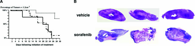
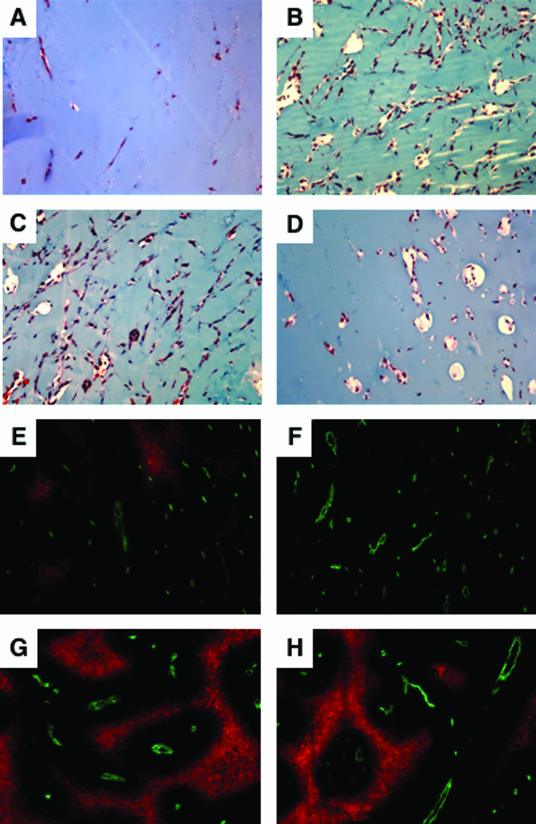
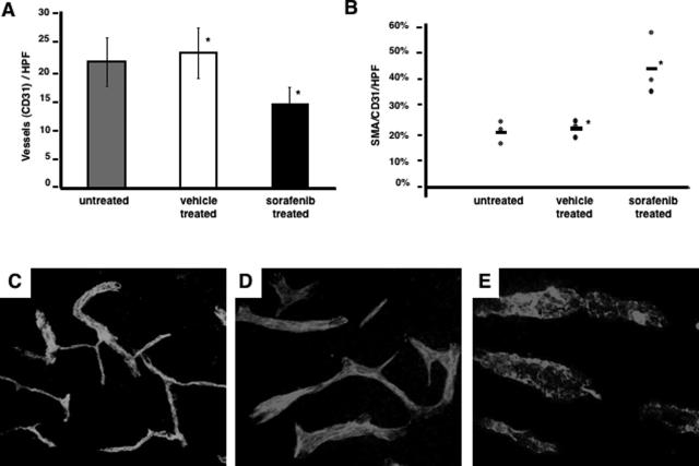
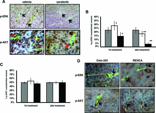
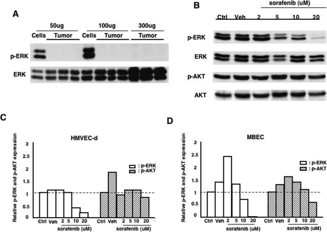
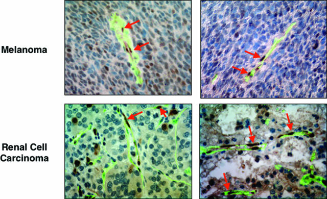
Activation of the Raf-MEK-ERK signal transduction pathway in endothelial cells is required for angiogenesis. Raf is the kinase most efficiently inhibited by the multikinase inhibitor sorafenib, which has shown activity against certain human cancers in clinical trials. To understand the mechanisms underlying this activity, we studied how it controlled growth of K1735 murine melanomas. Therapy caused massive regional tumor cell death accompanied by severe tumor hypoxia, decreased microvessel density, increased percentage of pericyte-covered vessels, and increased caliber and decreased arborization of vessels. These signs of K1735 angiogenesis inhibition, along with its ability to inhibit Matrigel neovascularization, showed that sorafenib is an effective anti-angiogenic agent. Extracellular signal-regulated kinase (ERK) activation in tumor endothelial cells, revealed by immunostaining for phospho-ERK and CD34, was inhibited, whereas AKT activation, revealed by phospho-AKT immunostaining, was not inhibited in K1735 and two other tumor types treated with sorafenib. Treatment decreased endothelial but not tumor cell proliferation and increased both endothelial cell and tumor cell apoptosis. These data indicate that sorafenib's anti-tumor efficacy may be primarily attributable to angiogenesis inhibition resulting from its inhibition of Raf-MEK-ERK signaling in endothelial cells. Assessing endothelial cell ERK activation in tumor bio-psies may provide mechanistic insights into and allow monitoring of sorafenib's activity in patients in clinical trials.


Figures

Figure 1
Figure 1
K1735 tumor growth is inhibited by sorafenib treatment. Female C3H/HeN mice bearing 1- to 2-mm diameter K1735 tumors were started on treatment with sorafenib (30 mg/kg) or vehicle by gavage daily. Tumor size was measure with calipers, and volumes were calculated using the formula 0.5× (width)2 × (length). Plotted is time taken by tumors to reach 1.2-cm3 volume at which time mice were euthanized (x axis, days after initiation of treatment; y axis, percentage of tumors in each treatment group <1.2 cm3). A: Untreated tumors are represented by black lines (n = 4), vehicle-treated tumors are represented by dashed lines (n = 9), and sorafenib-treated tumors are represented by dotted lines (n = 10). B: Sections from sorafenib or vehicle-treated K1735 tumors were stained with H&E. Pink regions are primarily necrotic; purple regions are primarily viable.
Figure 2
Figure 2
Effect of sorafenib on Matrigel neovascularization and tumor ischemia. Matrigel pellets containing PBS (A) or bFGF (100 ng/ml) (B–D) were implanted in mice that went untreated (A, B) or were treated with vehicle (C) or sorafenib (30 mg/kg) (D) for 7 days. Sections of the pellets were stained with Masson-Trichrome to reveal vascular structures containing erythrocytes (red). Mice with K1735 tumors were treated with vehicle or sorafenib (30 mg/kg) for 7 days. These mice were injected with EF5 to label hypoxic cells and fluorescein isothiocyanate-tomato lectin to illuminate perfused vessels before tumor excision. Shown are representative sections of vehicle-treated (E, F) and sorafenib-treated (G, H) tumors stained with Cy3-conjugated anti-EF5 monoclonal antibody. Red indicates severely hypoxic tumor regions labeled by EF5, and green indicates perfused tumor vessels. Original magnifications, ×100.
Figure 3
Figure 3
Inhibition of tumor angiogenesis by sorafenib. K1735 tumor vessels were revealed by CD31 staining and quantitated as previously described. A: Histograms of MVDs in untreated (n = 5, gray), vehicle-treated (n = 6, clear bar), and sorafenib-treated (n = 9, black bar) tumors are shown (*significant difference at P < 0.05, Student’s t-test). Tumor vessels were characterized for coverage by pericytes by staining for smooth muscle actin (SMA) in addition to CD31. B: The percentage of pericyte-covered vessels in individual untreated, vehicle-treated, and sorafenib-treated K1735 tumors are shown (n = 3). The mean of each group is indicated by a horizontal bar (*significant difference at P < 0.05, Student’s t-test). Thick sections of untreated (C), vehicle-treated (D), and sorafenib-treated (E) tumors were stained for CD31 and viewed by confocal microscopy. Original magnifications, ×200.
Figure 4
Figure 4
Inhibition of ERK but not AKT activation in tumor endothelial cells by sorafenib. K1735 tumors treated with either vehicle or sorafenib were stained with anti-p-ERK or anti-p-AKT antibody (brown immunohistochemistry) followed by vessel staining with anti-CD34 antibody (green immunofluorescence) and a light hematoxylin counterstain. A: The p-ERK/p-AKT images and the CD34 images of the same representative fields are superimposed; vascular ECs staining for p-ERK or p-AKT are indicated by arrows. Histograms of the percentage of tumor vessels staining for p-ERK (B) or p-AKT (C) are shown for tumors treated for 7 or 28 days with sorafenib (black bars; n = 3 for p-ERK, n = 6 for p-AKT) or vehicle (white bars; n = 3 for p-ERK, n = 4 for p-AKT) and for size-matched untreated tumors (gray bars, n = 3). *Indicates significant difference at P < 0.05; ** indicates significant difference at P < 0.01. D: Colo-205 and RENCA tumors were stained for p-ERK, p-AKT, and CD34 as described above. Original magnifications, ×400.
Figure 5
Figure 5
ERK and AKT activation in K1735 cells and tumors and the effect of sorafenib treatment on ERK and AKT activation in ECs. A: Western blots of lysates (50, 100, and 300 μg) of cultured K1735 tumor cells and K1735 tumors were probed for p-ERK and subsequently reprobed for ERK. Cultured HMVEC-d and MBECs were treated with vehicle or different concentrations of sorafenib for 2 hours. Western blots of cell lysates were probed for p-ERK and p-AKT. B: After stripping, these blots were reprobed for ERK and AKT, respectively. HMVEC-d expression of these antigens is shown. After densitometry of the relevant bands, p-ERK content was normalized for ERK content, and p-AKT content was normalized for AKT content. Shown are histograms of normalized p-ERK and p-AKT content for HMVEC-d (C) and MBECs (D) under different treatment conditions relative to normalized p-ERK and p-AKT content in untreated HMVEC-d and MBECs (set at 1.0).
Figure 6
Figure 6
ERK activation in human melanomas and renal cell carcinomas. Paraffin sections of human melanomas and renal cell carcinomas were stained with anti-p-ERK antibody (brown immunohistochemistry) followed by vessel staining with anti-CD34 antibody (green immunofluorescence) and a light hematoxylin counterstain. Tumor vascular endothelial cells expressing p-ERK are indicated by arrows. Original magnifications, ×400.

Similar articles

Cited by

References

    1. Almoguera C, Shibata D, Forrester K, Martin J, Arnheim N, Perucho M. Most human carcinomas of the exocrine pancreas contain mutant c-K-ras genes. Cell. 1988;53:549–554. - PubMed
    1. Forrester K, Almoguera C, Han K, Grizzle WE, Perucho M. Detection of high incidence of K-ras oncogenes during human colon tumorigenesis. Nature. 1987;327:298–303. - PubMed
    1. Bos JL, Fearon ER, Hamilton SR, Verlaan-de Vries M, van Boom JH, van der Eb AJ, Vogelstein B. Prevalence of ras gene mutations in human colorectal cancers. Nature. 1987;327:293–297. - PubMed
    1. Bos JL. Ras oncogenes in human cancer: a review. Cancer Res. 1989;49:4682–4689. - PubMed
    1. Brose MS, Volpe P, Feldman M, Kumar M, Rishi I, Gerrero R, Einhorn E, Herlyn M, Minna J, Nicholson A, Roth JA, Albelda SM, Davies H, Cox C, Brignell G, Stephens P, Futreal PA, Wooster R, Stratton MR, Weber BL. BRAF and RAS mutations in human lung cancer and melanoma. Cancer Res. 2002;62:6997–7000. - PubMed

Publication types

MeSH terms