. 2006;34(20):6023-33.
doi: 10.1093/nar/gkl744. Epub 2006 Oct 27.

Proliferating cell nuclear antigen loaded onto double-stranded DNA: dynamics, minor groove interactions and functional implications

Affiliations

Proliferating cell nuclear antigen loaded onto double-stranded DNA: dynamics, minor groove interactions and functional implications

Ivaylo Ivanov et al. Nucleic Acids Res. 2006.

Abstract

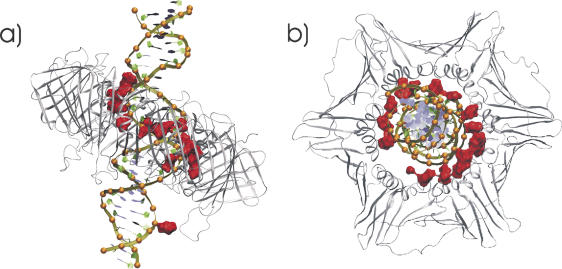
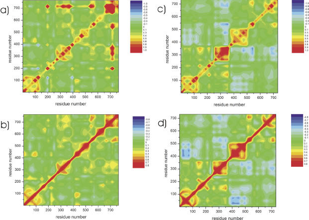
Proliferating cell nuclear antigen (PCNA) acts as a biologically essential processivity factor that encircles DNA and provides binding sites for polymerase, flap endonuclease-1 (FEN-1) and ligase during DNA replication and repair. We have computationally characterized the interactions of human and Archaeoglobus fulgidus PCNA trimer with double-stranded DNA (ds DNA) using multi-nanosecond classical molecular dynamics simulations. The results reveal the interactions of DNA passing through the PCNA trimeric ring including the contacts formed, overall orientation and motion with respect to the sliding clamp. Notably, we observe pronounced tilting of the axis of dsDNA with respect to the PCNA ring plane reflecting interactions between the DNA phosphodiester backbone and positively charged arginine and lysine residues lining the PCNA inner surface. Covariance matrix analysis revealed a pattern of correlated motions within and between the three equivalent subunits involving the PCNA C-terminal region and linker strand associated with partner protein binding sites. Additionally, principal component analysis identified low frequency global PCNA subunit motions suitable for translocation along duplex DNA. The PCNA motions and interactions with the DNA minor groove, identified here computationally, provide an unexpected basis for PCNA to act in the coordinated handoff of intermediates from polymerase to FEN-1 to ligase during DNA replication and repair.


Figures

Figure 1
Figure 1
hPCNA top and side view of the final configuration from molecular dynamics. Color-coding scheme according to secondary structure.
Figure 2
Figure 2
PCNA interactions with dsDNA: a) side view and b) upper view of the hPCNA dsDNA complex reveal the relationship between the tilting of the hPCNA ring with respect to dsDNA and the interactions between the nucleic acid backbone and the inner positively charged surface of the sliding clamp. The dsDNA phosphodiester groups (orange spheres) plus basic (arginine and lysine) groups of hPCNA within 6 Å of the phosphates (red surfaces) are shown.
Figure 3
Figure 3
Histogram of the interactions between the side chain N atoms of basic (arginine and lysine) groups of PCNA located within 7 Å of the nucleic acid backbone P atoms. The distribution of nitrogen–phosphorus contacts as a function of distance reveals two peaks with a minimum at ∼4.6 Å. Thus, the N–P contacts can be naturally subdivided into close (below the minimum) and distant (above the minimum) interactions. This is true for both aPCNA (blue) and hPCNA (green). The red curve represents the weighting function that was used to select ‘close’ interactions. It varies between 0 and 1 and for clarity is not shown to scale.
Figure 4
Figure 4
Time evolution of the interactions between the side chain N atoms of basic (arginine and lysine) groups of hPCNA located within 7 Å of the nucleic acid backbone P atoms. Data for aPCNA is shown on (a) and for hPCNA on (b). (c and d) display the time evolution of the tilt angle between the DNA axis and the plane of the PCNA ring for aPCNA and hPCNA, respectively. The observed increase in the number of N–P contacts over the trajectory is closely paralleled by an increase in the tilt angle, which approaches 20° toward the end of the simulations.
Figure 5
Figure 5
Time evolution of ‘close’ nitrogen–phosphorus contacts (within a 4.6 Å cutoff) between each of the PCNA subunits and the nucleic acid. Data for aPCNA is shown on (a) and for hPCNA on (b), respectively. Individual subunit graphs are highlighted in red, green and blue. The figure essentially represents the share of P–N contacts with the DNA phosphodiester backbone made by each of the PCNA subunits during the simulation, highlighting the dynamic nature of the observed asymmetric binding between the PCNA subunits and DNA.
Figure 6
Figure 6
Computed versus experimental B-factors identifying the mobile regions of the PCNA trimer as a function of residue number (with subunits A, B and C shown sequentially): (a) for hPCNA and (b) for aPCNA.
Figure 7
Figure 7
Correlated and anti-correlated PCNA motions revealed by cross-correlation and covariance maps for hPCNA and aPCNA. a) Covariance (a) and cross-correlation matrix (b) for hPCNA; b) Covariance (c) and cross-correlation matrix (d) for aPCNA. The cross-correlation and covariance maps identify regions of the protein that may be distant in the sequence but, nevertheless, move in a concerted way. Positive values (red) denote correlated motions; negative values (blue) indicated anti-correlated motions.
Figure 8
Figure 8
Front (a) and side (b) representations of the first principal component vector mapped onto the Cα carbon atoms of hPCNA. The three subunits are shown in trace representation along the Cα carbon atoms in red, green and blue, respectively.
Figure 9
Figure 9
Histograms obtained from two-dimensional projections of the trajectories onto the first two principal eigenvectors. Data for hPCNA and aPCNA is shown in (a) and (b), respectively.

Similar articles

Cited by

References

    1. Krishna T.S., Kong X.P., Gary S., Burgers P.M., Kuriyan J. Crystal structure of the eukaryotic DNA polymerase processivity factor PCNA. Cell. 1994;79:1233–1243. - PubMed
    1. Kelman Z., O'Donnell M. Structural and functional similarities of prokaryotic and eukaryotic DNA polymerase sliding clamps. Nucleic Acids Res. 1995;23:3613–3620. - PMC - PubMed
    1. Gulbis J.M., Kelman Z., Hurwitz J., O'Donnell M., Kuriyan J. Structure of the C-terminal region of p21(WAF1/CIP1) complexed with human PCNA. Cell. 1996;87:297–306. - PubMed
    1. Moarefi I., Jeruzalmi D., Turner J., O'Donnell M., Kuriyan J. Crystal structure of the DNA polymerase processivity factor of T4 bacteriophage. J. Mol. Biol. 2000;296:1215–1223. - PubMed
    1. Maga G., Hubscher U. Proliferating cell nuclear antigen (PCNA): a dancer with many partners. J. Cell. Sci. 2003;116:3051–3060. - PubMed

Publication types