. 2007 Feb;292(2):L519-28.
doi: 10.1152/ajplung.00312.2006. Epub 2006 Oct 27.

IL-23 mediates inflammatory responses to mucoid Pseudomonas aeruginosa lung infection in mice

Affiliations

IL-23 mediates inflammatory responses to mucoid Pseudomonas aeruginosa lung infection in mice

Patricia J Dubin et al. Am J Physiol Lung Cell Mol Physiol. 2007 Feb.

Abstract

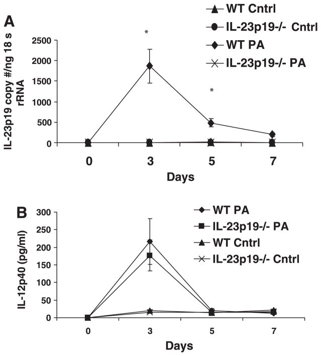
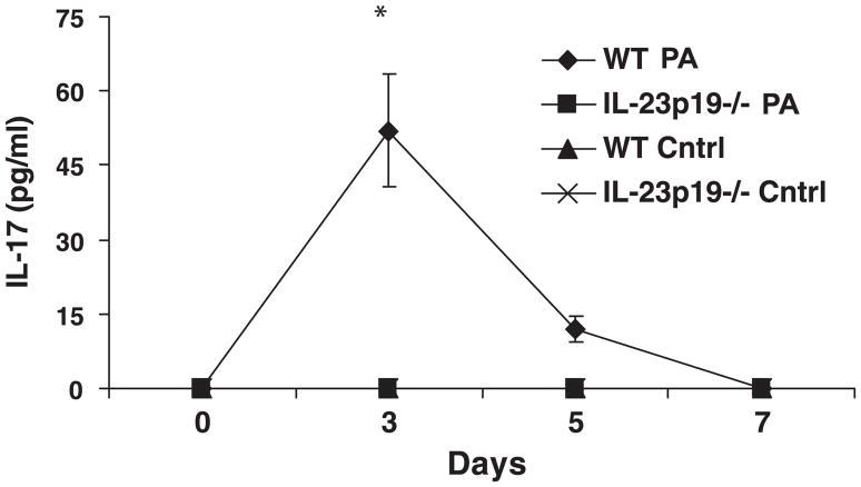
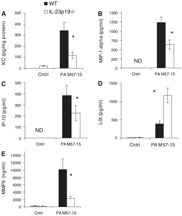
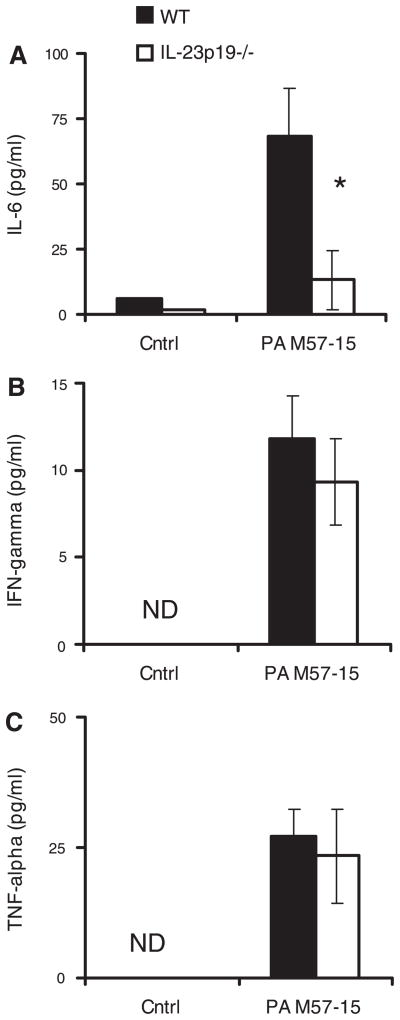
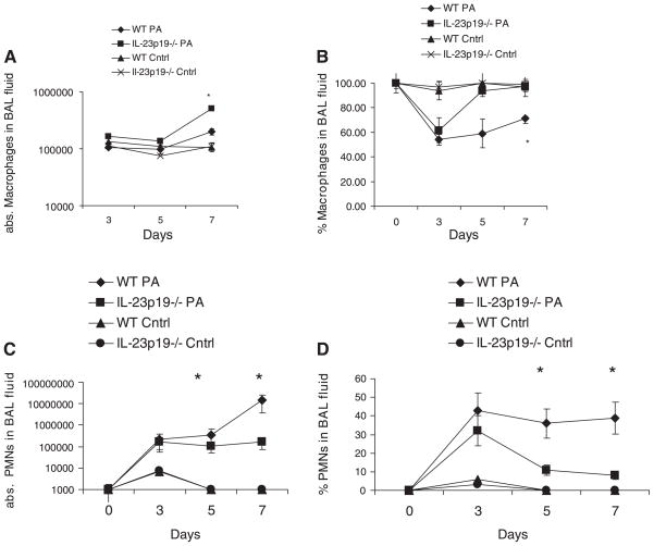
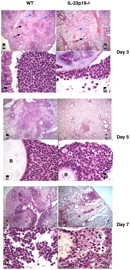
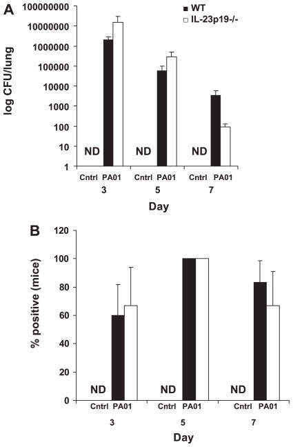
Patients with cystic fibrosis (CF) develop chronic Pseudomonas aeruginosa lung infection with mucoid strains of P. aeruginosa; these infections cause significant morbidity. The immunological response in these infections is characterized by an influx of neutrophils to the lung and subsequent lung damage over time; however, the underlying mediators to this response are not well understood. We recently reported that IL-23 and IL-17 were elevated in the sputum of patients with CF who were actively infected with P. aeruginosa; however, the importance of IL-23 and IL-17 in mediating this inflammation was unclear. To understand the role that IL-23 plays in initiating airway inflammation in response to P. aeruginosa, IL-23p19(-/-) (IL-23 deficient) and wild-type (WT) mice were challenged with agarose beads containing a clinical, mucoid isolate of P. aeruginosa. Levels of proinflammatory cytokines, chemokines, bacterial dissemination, and inflammatory infiltrates were measured. IL-23-deficient mice had significantly lower induction of IL-17, keratinocyte-derived chemokine (KC), and IL-6, decreased bronchoalveolar lavage (BAL) neutrophils, metalloproteinase-9 (MMP-9), and reduced airway inflammation than WT mice. Despite the reduced level of inflammation in IL-23p19(-/-) mice, there were no differences in the induction of TNF and interferon-gamma or in bacterial dissemination between the two groups. This study demonstrates that IL-23 plays a critical role in generating airway inflammation observed in mucoid P. aeruginosa infection and suggests that IL-23 could be a potential target for immunotherapy to treat airway inflammation in CF.


Figures

Fig. 1
Fig. 1
A: expression of IL-23p19 mRNA measured by TaqMan real-time PCR. Mice were killed after being infected with Pseudomonas aeruginosa strain PA M57–15-laden agarose beads for 0, 3, 5, and 7 days or uninfected control beads (Cntrl). IL-23p19−/− exhibited no detectable IL-23p19, whereas the wild-type (WT) infected mice had significantly increased IL-23p19 expression at days 3 and 5. There was no detectable IL-23p19 production in control mice. B: IL-12p40 was measured in the bronchoalveolar lavage (BAL) fluid at the protein level. There were no significant differences between infected WT and IL-23p19−/− mice. There is detectable production at baseline and for control mice of both groups and noted increase in IL-12p40 production in both WT and IL-23p19−/− mice at day 3. N = 3–5 per group for A and B.*P < 0.05.
Fig. 2
Fig. 2
IL-17 protein measured by ELISA in BAL. IL-17 was measured at 0, 3, 5, and 7 days post-infection (n = 5 per group); *P < 0.05. IL-23p19−/− mice exhibited no detectable IL-17, whereas the WT infected mice had significantly increased IL-17 expression at day 3. IL-17 remained elevated at day 5 in WT infected mice but was not significantly greater than the other groups. There was no detectable production of IL-17 in the WT or IL-23p19−/− control mice.
Fig. 3
Fig. 3
Chemokine and metalloproteinase (MMP) levels in lung homogenate and BAL fluid during chronic P. aeruginosa infection. Keratinocyte-derived chemokine (KC) (A) and MMP-9 (E) from lung homogenate were measured by LINCOplex and reported for day 5 after infection (n = 3–5 per group); *P < 0.05. LPS-induced CXC chemokine (LIX) (D) from BAL was measured by ELISA and reported for day 5 after infection (n = 5); *P < 0.05. Macrophage inflammatory protein-1α (MIP-1α) (B) and IFN-γ-inducible protein 10 (IP-10) (C) from BAL were measured by LINCOplex and reported for day 5 after infection. (n = 5); *P < 0.05. Levels in the mice treated with control beads were either not detectable (ND) or not significantly different from baseline. In addition, WT controls were not significantly different from IL-23p19−/− controls.
Fig. 4
Fig. 4
Cytokine levels in BAL fluid during chronic P. aeruginosa infection. IL-6 (A), IFN-γ (D), and TNF-α (C), measured by Luminex, are reported for day 5 after infection. IL-6 was significantly increased in infected WT mice compared with IL-23p19−/− infected mice. Infected WT mice had significantly elevated levels over control WT mice. There was no significant difference between WT and IL-23p19−/− control mice (n = 5 per group); *P < 0.05.
Fig. 5
Fig. 5
Cellular recruitment during chronic P. aeruginosa infection. A: absolute (abs.) numbers of recovered macrophages in BAL fluid from WT and IL-23p19−/− mice on days 0, 3, 5, and 7. B: percent macrophages in BAL fluid from WT and IL-23p19−/− on days 0, 3, 5, and 7 after infection. C: absolute numbers of recovered neutrophils (PMNs) in BAL fluid from WT and IL-23p19−/− mice on days 0, 3, 5, and 7. D: percent neutrophils in BAL fluid from WT and IL-23p19−/− mice on days 0, 3, 5, and 7 after infection. Where significant differences are noted, the infected WT mice demonstrate higher levels than the IL-23p19−/− infected mice and the WT control mice; there is no significant difference between WT and IL-23p19−/− control mice (n = 10 per group); *P < 0.05.
Fig. 6
Fig. 6
Representative photomicrographs of lung from WT and IL-23p19−/− mice infected with PA M57–15-laden beads at days 3, 5, and 7. Top panels for each time point are at ×50 magnification, and bottom panels are at ×1,000 magnification. Day 3 (ad): black arrows indicate airways with marked inflammation, and blue arrows indicate beads. Day 5 (eh): B marks agarose bead in the airway lumen. il: day 7.
Fig. 7
Fig. 7
Quantitative scoring of intraluminal (A) and peribronchial (B) infiltrate: comparing WT and IL-23p19−/− infected and control demonstrates significant differences at days 3, 5, and 7 for the infected WT mice compared with the IL-23p19−/− infected and WT control groups (n = 5 per group); *P < 0.05.
Fig. 8
Fig. 8
Quantitative comparisons of bacterial burden in the lung by comparison of colony-forming units (CFU) (A) and percent of mice originally infected from which bacteria are recovered (B) (n = per group).

Similar articles

Cited by

References

    1. Aggarwal S, Ghilardi N, Xie MH, de Sauvage FJ, Gurney AL. Interleukin-23 promotes a distinct CD4 T cell activation state characterized by the production of interleukin-17. J Biol Chem. 2003;278:1910–1914. - PubMed
    1. Armstrong DS, Hook SM, Jamsen KM, Nixon GM, Carzino R, Carlin JB, Robertson CF, Grimwood K. Lower airway inflammation in infants with cystic fibrosis detected by newborn screening. Pediatr Pulmonol. 2005;40:500–510. - PubMed
    1. Bonfield TL, Panuska JR, Konstan MW, Hilliard KA, Hilliard JB, Ghnaim H, Berger M. Inflammatory cytokines in cystic fibrosis lungs. Am J Respir Crit Care Med. 1995;152:2111–2118. - PubMed
    1. Brauner A, Cryz SJ, Granstrom M, Hanson HS, Lofstrand L, Strandvik B, Wretlind B. Immunoglobulin G antibodies to Pseudomonas aeruginosa lipopolysaccharides and exotoxin A in patients with cystic fibrosis or bacteremia. Eur J Clin Microbiol Infect Dis. 1993;12:430–436. - PubMed
    1. Carpagnano GE, Barnes PJ, Geddes DM, Hodson ME, Kharitonov SA. Increased leukotriene B4 and interleukin-6 in exhaled breath condensate in cystic fibrosis. Am J Respir Crit Care Med. 2003;167:1109–1112. - PubMed

Publication types

MeSH terms

LinkOut - more resources