Atherosclerosis-related molecular alteration of the human CaV1.2 calcium channel alpha1C subunit
- PMID: 17071743
- PMCID: PMC1636572
- DOI: 10.1073/pnas.0606539103
Atherosclerosis-related molecular alteration of the human CaV1.2 calcium channel alpha1C subunit
Abstract
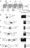
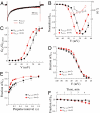
Atherosclerosis is an inflammatory process characterized by proliferation and dedifferentiation of vascular smooth muscle cells (VSMC). Ca(v)1.2 calcium channels may have a role in atherosclerosis because they are essential for Ca(2+)-signal transduction in VSMC. The pore-forming Ca(v)1.2alpha1 subunit of the channel is subject to alternative splicing. Here, we investigated whether the Ca(v)1.2alpha1 splice variants are affected by atherosclerosis. VSMC were isolated by laser-capture microdissection from frozen sections of adjacent regions of arteries affected and not affected by atherosclerosis. In VSMC from nonatherosclerotic regions, RT-PCR analysis revealed an extended repertoire of Ca(v)1.2alpha1 transcripts characterized by the presence of exons 21 and 41A. In VSMC affected by atherosclerosis, expression of the Ca(v)1.2alpha1 transcript was reduced and the Ca(v)1.2alpha1 splice variants were replaced with the unique exon-22 isoform lacking exon 41A. Molecular remodeling of the Ca(v)1.2alpha1 subunits associated with atherosclerosis caused changes in electrophysiological properties of the channels, including the kinetics and voltage-dependence of inactivation, recovery from inactivation, and rundown of the Ca(2+) current. Consistent with the pathophysiological state of VSMC in atherosclerosis, cell culture data pointed to a potentially important association of the exon-22 isoform of Ca(v)1.2alpha1 with proliferation of VSMC. Our findings are consistent with a hypothesis that localized changes in cytokine expression generated by inflammation in atherosclerosis affect alternative splicing of the Ca(v)1.2alpha1 gene in the human artery that causes molecular and electrophysiological remodeling of Ca(v)1.2 calcium channels and possibly affects VSMC proliferation.
Conflict of interest statement
The authors declare no conflict of interest.
Figures


Similar articles
-
Alternative splicing of Cav1.2 channel exons in smooth muscle cells of resistance-size arteries generates currents with unique electrophysiological properties.Am J Physiol Heart Circ Physiol. 2009 Aug;297(2):H680-8. doi: 10.1152/ajpheart.00109.2009. Epub 2009 Jun 5. Am J Physiol Heart Circ Physiol. 2009. PMID: 19502562 Free PMC article.
-
Gene splicing of an invertebrate beta subunit (LCavβ) in the N-terminal and HOOK domains and its regulation of LCav1 and LCav2 calcium channels.PLoS One. 2014 Apr 1;9(4):e92941. doi: 10.1371/journal.pone.0092941. eCollection 2014. PLoS One. 2014. PMID: 24690951 Free PMC article.
-
Smooth muscle-selective alternatively spliced exon generates functional variation in Cav1.2 calcium channels.J Biol Chem. 2004 Nov 26;279(48):50329-35. doi: 10.1074/jbc.M409436200. Epub 2004 Sep 20. J Biol Chem. 2004. PMID: 15381693
-
L-type CaV1.2 calcium channels: from in vitro findings to in vivo function.Physiol Rev. 2014 Jan;94(1):303-26. doi: 10.1152/physrev.00016.2013. Physiol Rev. 2014. PMID: 24382889 Review.
-
Splicing for alternative structures of Cav1.2 Ca2+ channels in cardiac and smooth muscles.Cardiovasc Res. 2005 Nov 1;68(2):197-203. doi: 10.1016/j.cardiores.2005.06.024. Epub 2005 Jul 27. Cardiovasc Res. 2005. PMID: 16051206 Review.
Cited by
-
Molecular alteration of Ca(v)1.2 calcium channel in chronic myocardial infarction.Pflugers Arch. 2009 Aug;458(4):701-11. doi: 10.1007/s00424-009-0652-4. Epub 2009 Mar 5. Pflugers Arch. 2009. PMID: 19263075
-
Aberrant Splicing Promotes Proteasomal Degradation of L-type CaV1.2 Calcium Channels by Competitive Binding for CaVβ Subunits in Cardiac Hypertrophy.Sci Rep. 2016 Oct 12;6:35247. doi: 10.1038/srep35247. Sci Rep. 2016. PMID: 27731386 Free PMC article.
-
Contribution of transient and sustained calcium influx, and sensitization to depolarization-induced contractions of the intact mouse aorta.BMC Physiol. 2012 Sep 3;12:9. doi: 10.1186/1472-6793-12-9. BMC Physiol. 2012. PMID: 22943445 Free PMC article.
-
CACNA1C (Cav1.2) in the pathophysiology of psychiatric disease.Prog Neurobiol. 2012 Oct;99(1):1-14. doi: 10.1016/j.pneurobio.2012.06.001. Epub 2012 Jun 15. Prog Neurobiol. 2012. PMID: 22705413 Free PMC article. Review.
-
Greater efficacy of atorvastatin versus a non-statin lipid-lowering agent against renal injury: potential role as a histone deacetylase inhibitor.Sci Rep. 2016 Nov 30;6:38034. doi: 10.1038/srep38034. Sci Rep. 2016. PMID: 27901066 Free PMC article.
References
-
- Ross R. N Engl J Med. 1999;340:115–126. - PubMed
-
- Abernethy DR, Schwartz JB. N Engl J Med. 1999;341:1447–1457. - PubMed
-
- Catterall WA. Annu Rev Cell Dev Biol. 2000;16:521–555. - PubMed
-
- Striessnig J. Cell Physiol Biochem. 1999;9:242–269. - PubMed
-
- Hu H, Marban E. Mol Pharmacol. 1998;53:902–907. - PubMed
Publication types
MeSH terms
Substances
Associated data
- Actions
- Actions
- Actions
- Actions
- Actions
Grants and funding
LinkOut - more resources
Full Text Sources
Other Literature Sources
Medical
Molecular Biology Databases
Miscellaneous

