. 2007 Feb;189(4):1266-78.
doi: 10.1128/JB.01130-06. Epub 2006 Oct 27.

The yefM-yoeB toxin-antitoxin systems of Escherichia coli and Streptococcus pneumoniae: functional and structural correlation

Affiliations

The yefM-yoeB toxin-antitoxin systems of Escherichia coli and Streptococcus pneumoniae: functional and structural correlation

Concha Nieto et al. J Bacteriol. 2007 Feb.

Abstract

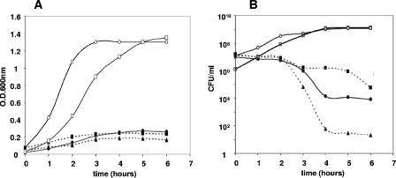
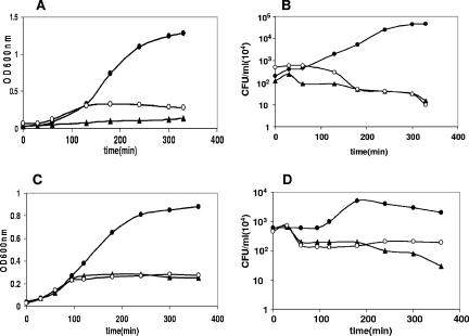
Toxin-antitoxin loci belonging to the yefM-yoeB family are located in the chromosome or in some plasmids of several bacteria. We cloned the yefM-yoeB locus of Streptococcus pneumoniae, and these genes encode bona fide antitoxin (YefM(Spn)) and toxin (YoeB(Spn)) products. We showed that overproduction of YoeB(Spn) is toxic to Escherichia coli cells, leading to severe inhibition of cell growth and to a reduction in cell viability; this toxicity was more pronounced in an E. coli B strain than in two E. coli K-12 strains. The YoeB(Spn)-mediated toxicity could be reversed by the cognate antitoxin, YefM(Spn), but not by overproduction of the E. coli YefM antitoxin. The pneumococcal proteins were purified and were shown to interact with each other both in vitro and in vivo. Far-UV circular dichroism analyses indicated that the pneumococcal antitoxin was partially, but not totally, unfolded and was different than its E. coli counterpart. Molecular modeling showed that the toxins belonging to the family were homologous, whereas the antitoxins appeared to be specifically designed for each bacterial locus; thus, the toxin-antitoxin interactions were adapted to the different bacterial environmental conditions. Both structural features, folding and the molecular modeled structure, could explain the lack of cross-complementation between the pneumococcal and E. coli antitoxins.


Figures

FIG. 1.
FIG. 1.
Effect of yoeBSpn expression in E. coli strains. (A) Cell growth arrest after induction of yoeBSpn expression in strains MG1655 (○ and •), TOP-10 (▪), and BL21 (▴) harboring pYBS2 (yoeBSpn). Cells were grown exponentially in medium containing 0.2% glucose (repression conditions) and kanamycin to an OD600 of 0.03 to 0.04. Cultures were divided in two. One half of each culture was grown in the presence of glucose (open symbols; for clarity, only the data for strain MG1655 are shown), and the other half was induced by addition of 0.2% arabinose (solid symbols). Growth was monitored by determining the OD600 of the cultures. (B) At the times indicated, the numbers of CFU in the cultures were determined by plating appropriate dilutions on TY agar supplemented with 0.4% glucose and kanamycin and incubating the preparations overnight at 37°C. Neither cell growth arrest nor a reduction in the number of CFU was observed for E. coli TOP-10 cultures containing the pYFS2 plasmid (YefMSpn) (□) after induction of antitoxin synthesis by addition of 2 mM IPTG. This culture was grown and plated as described above but using ampicillin instead of kanamycin for selection. All experiments were performed at least in duplicate.
FIG. 2.
FIG. 2.
Toxicity of YoeBSpn is neutralized only by combined expression of the cognate antitoxin in E. coli. (A and B) E. coli TOP-10 strains harboring pYBS2 (yoeBSpn) (○), pYBS2 (yoeBSpn), pYFS10 (yefMSpn) (•), or pYBS2 (yoeBSpn) and pNMYE (yefMEco) (▴) were grown exponentially in TY medium containing 0.2% glucose, kanamycin, and ampicillin to an OD600 of 0.03 to 0.04. Then 0.2% arabinose (toxin synthesis induction) and 1 mM IPTG (antitoxin synthesis induction) were added. (A) Absorbance at 600 nm was used to monitor the growth of the cultures. (B) At the times indicated, samples were diluted and plated on TY agar containing 0.4% glucose, kanamycin, and ampicillin and incubated overnight at 37°C. (C and D) E. coli TOP-10 strains harboring pFYBE (yoeBEco) (○), pFYBE (yoeBEco) and pNMYE (yefMEco) (•), or pFYBE (yoeBEco) and pYFS10 (yefMSpn) (▴) were grown and treated as described above. (C) Absorbance at 600 nm was used to monitor the growth of the cultures. (D) At the times indicated, samples were diluted and plated on TY agar containing 0.4% glucose, kanamycin, and ampicillin and incubated overnight at 37°C. All the experiments were performed at least in duplicate.
FIG. 3.
FIG. 3.
Recovery of cell viability due to antitoxin synthesis after overproduction of YoeBSpn. E. coli TOP-10 cells harboring plasmids pYBS2 (yoeBSpn) and pYFS10 (yefMSpn) were grown in TY medium without glucose containing kanamycin and ampicillin. When the culture reached an OD600 of 0.13, 0.2% arabinose was added. Absorbance data were obtained for 7 h after induction of toxin synthesis (inset). At different times, samples were plated on TY agar containing 0.4% glucose, kanamycin, and ampicillin supplemented (•) or not supplemented (○) with IPTG.
FIG. 4.
FIG. 4.
Differences in YoeBSpn and RelE2Spn toxicity in E. coli lon+ and lon strains. (A) Inhibition of cell growth by overproduction of the pneumococcal toxins YoeBSpn (○ and •) and RelE2Spn (▴ and ▵) in E. coli strain MG1655 (lon+) (solid symbols) or in the lon isogenic mutant MG1655lon (open symbols). E. coli MG1655 (• and ▴) or MG1655lon (○) was grown in TY medium supplemented with kanamycin and 0.2% glucose (▪) or arabinose (•, ○, ▴, and ▵). Absorbance data were obtained at different times after the addition of arabinose. (B) To determine the number of CFU, samples were removed at various times and spread on TY agar plates containing 0.4% glucose and kanamycin. The percentages of viable cells were calculated by comparison of the numbers of CFU in the induced cultures to the numbers of CFU in the noninduced cultures at time zero.
FIG. 5.
FIG. 5.
Purification of (YefM-YoeB-His)Spn complex and His-YefMSpn antitoxin and indications of YefM-YoeBSpn interaction. (A and B) Chromatograms showing the elution pattern of proteins from the nickel affinity column. Note that although the yield of the YefMSpn protein (B) was poor, an increase in the absorbance was evident. However, no peak was observed, most likely because of the small amount of protein which was masked by the increase in the imidazole background absorbance. The results of SDS-PAGE analyses of eluted fractions are shown in the insets. RT, retention time; mAu, milli-absorbance units. (C) Elution profile of the YefM-YoeBSpn protein complex from a 16/60 Sephacryl S-100 HR gel filtration column, showing a void volume of 45.89. The 45.89-ml peak with a Kav value of 0.1351 corresponds to an Mr of approximately 49,500. (D) E. coli two-hybrid indicator strain (Stratagene) (derived from XL-1 Blue MRF′) harboring plasmids pBT-LGF2 and pTRG-Gal11P (positive control) (streak 1), pBT-yefMSpn and pTRG (streak 2), pBT and pTRG-yoeBSpn (streak 3), pBT and pTRG-Gal11P (streak 4), pBT-yefMSpn and pTRG-yoeBSpn (streak 5), or pBT-yoeBSpn and pTRG-yefMSpn (streak 6) was streaked on selective medium containing 3-AT and incubated overnight at 37°C. Cotransformants in streaks 2, 3, and 4 were used as negative controls.
FIG. 6.
FIG. 6.
Far-UV CD spectra of His-YefMSpn (A) and (YefM-YoeB-His)Spn complex (B) at 4°C (solid line), 37°C (dashed line), and 60°C (dotted line). (C) CD ellipticity of His-YefMSpn (▴) and (YefM-YoeB-His)Spn (○) was measured at 220 nm at various temperatures to estimate the thermal stability. The values were normalized between 0 and 1 and are expressed as fractional changes. (D) SDS-PAGE analysis of (YefM-YoeB-His)Spn sample that was examined by CD analysis before and after exposure to 85°C. Lane M, marker; lane 1, before treatment (sample at room temperature); lane 2, after treatment (soluble fraction after exposure to 85°C).
FIG. 7.
FIG. 7.
Surface electrostatic potentials of the interaction domains of the antitoxins YefMEco (A), YefMSpn model (C), and Axe model (E) with their cognate YoeB toxins and surface electrostatic potentials of the toxins YoeBEco (B), YoeBSpn model (D), and Txe model (F). All the surfaces are oriented facing the interacting side and in equivalent positions (by superposition with the CE algorithm). Positive potentials are blue, and negative potentials are red.

References

    1. Aizenman, E., H. Engelberg-Kulka, and G. Glaser. 1996. An Escherichia coli chromosomal “addiction module” regulated by guanosine 3′,5′-bispyrophosphate: a model for programmed bacterial cell death. Proc. Natl. Acad. Sci. USA 93:6059-6063. - PMC - PubMed
    1. Andrade, M. A., P. Chacón, J. J. Merolo, and F. Morán. 1993. Evaluation of secondary structure of proteins from UV circular dichroism spectra using an unsupervised learning neural network. Protein Eng. 6:443-454. - PubMed
    1. Baquero, F. 1996. Epidemiology and management of penicillin-resistant pneumococci. Curr. Opin. Infect. Dis. 9:372-379.
    1. Bravo, A., G. de Torrontegui, and R. Díaz. 1987. Identification of components of a new stability system of plasmid R1, Par D, that is close to the origin of replication of this plasmid. Mol. Gen. Genet. 210:101-110. - PubMed
    1. Cherny, I., and E. Gazit. 2004. The YefM antitoxin defines a family of natively unfolded proteins: implications as a novel antibacterial target. J. Biol. Chem. 279:8252-8261. - PubMed

Publication types

MeSH terms

LinkOut - more resources