Comparative Study
. 2007 Jan;189(2):388-402.
doi: 10.1128/JB.00981-06. Epub 2006 Oct 27.

Regulation of rugosity and biofilm formation in Vibrio cholerae: comparison of VpsT and VpsR regulons and epistasis analysis of vpsT, vpsR, and hapR

Affiliations
Comparative Study

Regulation of rugosity and biofilm formation in Vibrio cholerae: comparison of VpsT and VpsR regulons and epistasis analysis of vpsT, vpsR, and hapR

Sinem Beyhan et al. J Bacteriol. 2007 Jan.

Abstract

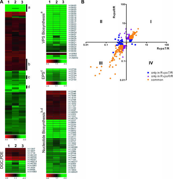
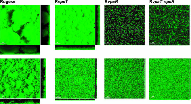
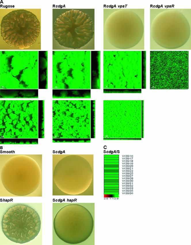
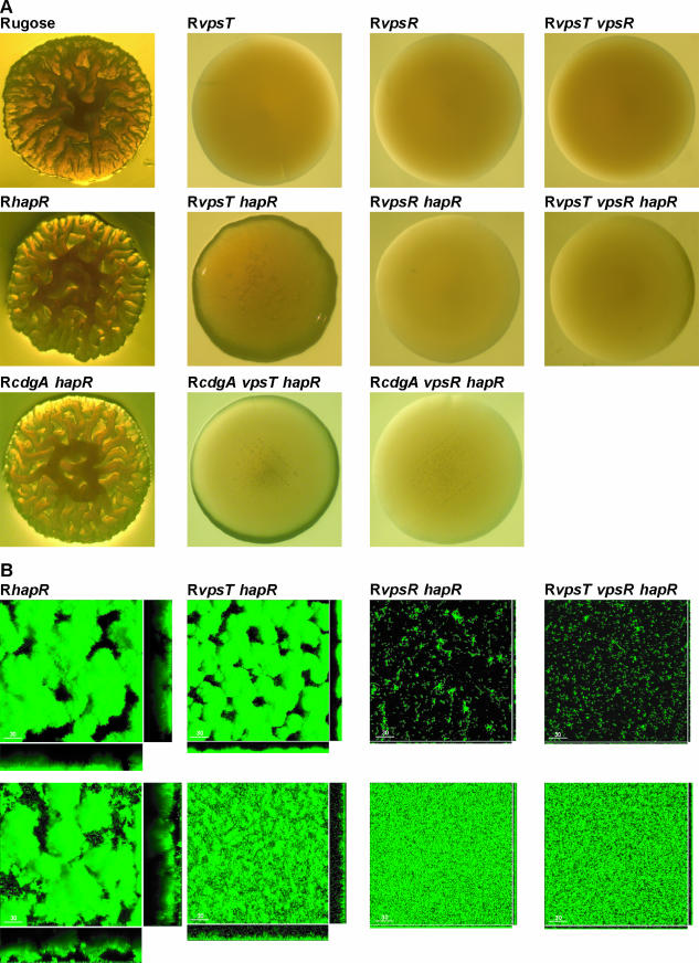
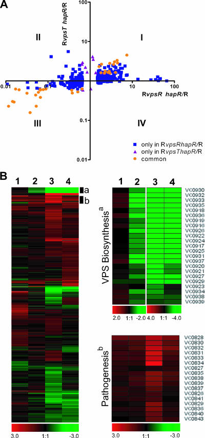
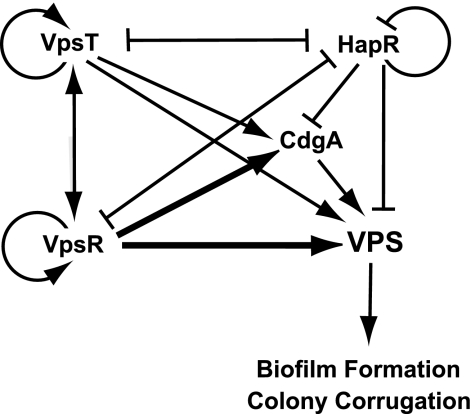
Vibrio cholerae undergoes phenotypic variation that generates two morphologically different variants, termed smooth and rugose. The transcriptional profiles of the two variants differ greatly, and many of the differentially regulated genes are controlled by a complex regulatory circuitry that includes the transcriptional regulators VpsR, VpsT, and HapR. In this study, we identified the VpsT regulon and compared the VpsT and VpsR regulons to elucidate the contribution of each positive regulator to the rugose variant transcriptional profile and associated phenotypes. We have found that although the VpsT and VpsR regulons are very similar, the magnitude of the gene regulation accomplished by each regulator is different. We also determined that cdgA, which encodes a GGDEF domain protein, is partially responsible for the altered vps gene expression between the vpsT and vpsR mutants. Analysis of epistatic relationships among hapR, vpsT, and vpsR with respect to a whole-genome expression profile, colony morphology, and biofilm formation revealed that vpsR is epistatic to hapR and vpsT. Expression of virulence genes was increased in a vpsR hapR double mutant relative to a hapR mutant, suggesting that VpsR negatively regulates virulence gene expression in the hapR mutant. These results show that a complex regulatory interplay among VpsT, VpsR, HapR, and GGDEF/EAL family proteins controls transcription of the genes required for Vibrio polysaccharide and virulence factor production in V. cholerae.


Figures

FIG. 1.
FIG. 1.
Expression profiles of RvpsT, RvpsR, and RvpsT vpsR mutants. (A) Differentially expressed genes in RvpsR (lane 1), RvpsT (lane 2), and RvpsT vpsR (lane 3) mutants are clustered by using Euclidian distance and average linkage hierarchical clustering. A compact view of clustered genes is presented using the log2-based color scale shown at the bottom of the panels (red, induced; green, repressed). Clusters that contain the genes required for VPS biosynthesis, the EPS general secretion system, and nucleotide biosynthesis are marked with black bars and labeled with a, c, and b and d, respectively. The expression profiles of these genes and genes encoding diguanylate cyclases (DGC) and/or phosphodiesterases (PDE) are also shown as separate heat maps. (B) Comparison of gene expression profiles of the RvpsT and RvpsR mutants. The x and y axes indicate the relative changes in gene expression (n-fold) in the RvpsT and RvpsR mutants relative to the wild-type rugose strain, respectively, whereby each spot represents a gene that was differentially regulated by 1.5-fold in at least one experiment. The plot area is divided into four regions (I to IV) based on the expression patterns relative to the wild-type rugose strain. Blue squares indicate genes differentially expressed only in the RvpsT mutant, purple triangles indicate genes differentially expressed only in the RvpsR mutant, and orange circles indicate genes differentially expressed in both mutants.
FIG. 2.
FIG. 2.
Characterization of biofilm formation under flow and nonflow conditions. Three-dimensional biofilm structures of the wild-type rugose strain and the RvpsT, RvpsR, and RvpsT vpsR strains formed after 24 h and 8 h postinoculation in a once-through flow cell (top row) and nonflow systems (bottom row), respectively. Images were acquired by confocal laser scanning microscopy, and top-down views (large panes) and orthogonal views (side panels) of biofilms are shown.
None
Characterization of the role of CdgA on colony development, biofilm formation, and regulation of vps expression. (A) Colony morphology of rugose, RcdgA, RcdgA vpsT, and RcdgA vpsR strains grown on LB agar plates after 48 h of incubation at 30°C (top row). Three-dimensional biofilm structures of rugose, RcdgA, RcdgA vpsT, and RcdgA vpsR strains formed after 24 h postinoculation in once-through flow systems at high (second row) and low magnifications (third row) are shown. Images were acquired by confocal laser scanning microscopy and top-down views (large panels) and orthogonal views (side panels) of biofilms are shown. (B) Colony morphology of smooth, ScdgA, ShapR, and ScdgA hapR strains grown on LB agar plates after 48 h of incubation at 30°C. The expression profiles of vps genes in the ScdgA mutant relative to the smooth variant are presented using the log2-based color scale shown at the bottom of the panels (red, induced; green, repressed).
None
Characterization of colony morphology and biofilm formation. (A) Colony morphology of rugose, RvpsT, RvpsR, RvpsT vpsR, RhapR, RvpsT hapR, RvpsR hapR, RvpsT vpsR hapR, RcdgA hapR, RcdgA vpsT hapR, and RcdgA vpsR hapR strains grown on LB agar plates after 48 h of incubation at 30°C. (B) Three-dimensional biofilm structures of RhapR, RvpsT hapR, RvpsR hapR, and RvpsT vpsR hapR strains formed after 24 h and 8 h postinoculation in once-through flow cell (top row) and nonflow systems (bottom row), respectively, are shown. Images were acquired by confocal laser scanning microscopy and top-down views (large panes) and orthogonal views (side panels) of biofilms are shown.
FIG. 5.
FIG. 5.
Expression profiles of RhapR, RvpsR hapR, RvpsT hapR, and RvpsT vpsR hapR mutants. (A) Comparison of gene expression profiles of RvpsT hapR and RvpsR hapR mutants. The x and y axes denote the relative changes in gene expression (n-fold) in RvpsR hapR and RvpsT hapR mutants relative to the wild-type rugose strain, respectively, whereby each spot represents a gene that was differentially regulated 1.5-fold in at least one experiment. The plot area is divided into four regions (I to IV) based on the expression patterns in comparison to the rugose wild-type strain. Blue squares indicate genes differentially expressed only in the RvpsR hapR mutant, purple triangles indicate genes differentially expressed only in the RvpsT hapR mutant, and orange circles indicate genes differentially expressed in both mutants. (B) Differentially expressed genes in RhapR (lane 1), RvpsT hapR (lane 2), RvpsR hapR (lane 3), and RvpsT vpsR hapR (lane 4) mutants relative to the rugose variant are clustered by using Euclidian distance and average linkage hierarchical clustering. A compact view of clustered genes is presented using the log2-based color scale shown at the bottom of the panels (red, induced; green, repressed). Clusters that contain the genes required for VPS biosynthesis and pathogenesis are marked with black bars and labeled a and b, respectively. The expression profiles of these genes are also shown as separate heat maps. Expression profiles of genes required for VPS biosynthesis and virulence factor production are shown in clustered form in the enlarged views at right.
FIG. 6.
FIG. 6.
Expression of rugosity-associated genes is controlled by a complex regulatory circuitry. The expression of vps genes is positively controlled by the regulators VpsR and VpsT, but the magnitude of regulation by VpsR is greater (denoted by darker lines). In contrast, the expression levels of vps genes are negatively controlled by HapR. Expression of VpsR and VpsT is negatively controlled by HapR; similarly, VpsR and VpsT negatively control hapR message levels, suggesting a presence of a regulatory loop. The expression of vps genes is positively controlled by CdgA. Transcription of cdgA is positively regulated by VpsR and VpsT and negatively regulated by HapR.

Similar articles

Cited by

References

    1. Ali, A., J. A. Johnson, A. A. Franco, D. J. Metzger, T. D. Connell, J. G. Morris, Jr., and S. Sozhamannan. 2000. Mutations in the extracellular protein secretion pathway genes (eps) interfere with rugose polysaccharide production in and motility of Vibrio cholerae. Infect. Immun. 68:1967-1974. - PMC - PubMed
    1. Bao, Y., D. P. Lies, H. Fu, and G. P. Roberts. 1991. An improved Tn7-based system for the single-copy insertion of cloned genes into chromosomes of gram-negative bacteria. Gene 109:167-168. - PubMed
    1. Beyhan, S., A. D. Tischler, A. Camilli, and F. H. Yildiz. 2006. Transcriptome and phenotypic responses of Vibrio cholerae to increased cyclic di-GMP level. J. Bacteriol. 188:3600-3613. - PMC - PubMed
    1. Brombacher, E., A. Baratto, C. Dorel, and P. Landini. 2006. Gene expression regulation by the curli activator CsgD protein: modulation of cellulose biosynthesis and control of negative determinants for microbial adhesion. J. Bacteriol. 188:2027-2037. - PMC - PubMed
    1. Carroll, P. A., K. T. Tashima, M. B. Rogers, V. J. DiRita, and S. B. Calderwood. 1997. Phase variation in tcpH modulates expression of the ToxR regulon in Vibrio cholerae. Mol. Microbiol. 25:1099-1111. - PubMed

Publication types

MeSH terms

Substances

LinkOut - more resources