

Hepatic angiomyolipoma with trace amounts of fat: a case report and literature review
- PMID: 17071805
- PMCID: PMC1860516
- DOI: 10.1136/jcp.2005.027227
Hepatic angiomyolipoma with trace amounts of fat: a case report and literature review
Abstract
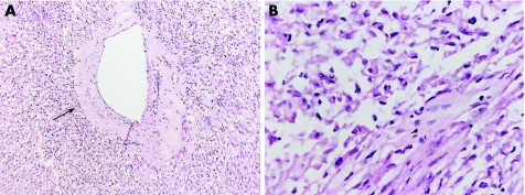
Hepatic angiomyolipoma (AML), a rare benign mesenchymal tumour, is characterised by the presence of mature adipose tissue, smooth-muscle cells and thick-walled blood vessels. Increasing attention to hepatic AMLs has led to the discovery that sufficient proportions of fat often allow for definite diagnoses preoperatively. However, the proportion of fatty tissue in these tumours is highly variable. One case of hepatic AML is reported, where the amount of fat was <1%. In this case, the viral hepatitis markers, including hepatitis B antigen and anti-hepatitis C virus antibody, were negative. The serum alpha-fetoprotein level was 3.4 ng/ml and in the normal range. Abdominal ultrasonography showed a hypoechoic mass measuring 5 cm in diameter and without an obvious capsule in the left lobe of the liver. A dynamic computed tomography scan showed a well-defined and slightly enhanced mass in the medial segment of the left lobe of the liver. Angiography showed that the mass was hypervascular in character. As hepatocellular carcinoma was highly suspected from these preoperative image studies, a left lobectomy was carried out. Microscopically, the amount of fat was too low to establish a diagnosis of hepatic AML. However, positive homatropine methylbromide 45 immunoreactivity of the smooth-muscle cells seemed to assist in arriving at the diagnosis.
Conflict of interest statement
Competing interests: None declared.


Similar articles
-
A misleading hepatic tumour: epithelioid angiomyolipoma.Acta Gastroenterol Belg. 2012 Dec;75(4):443-5. Acta Gastroenterol Belg. 2012. PMID: 23402089
-
Hepatic angiomyolipoma with a giant hemangioma.J Nippon Med Sch. 2011;78(5):317-21. doi: 10.1272/jnms.78.317. J Nippon Med Sch. 2011. PMID: 22041879
-
Angiomyolipoma of the liver with least amount of fat component: imaging features of CT, MR, and angiography.Abdom Imaging. 2002 Mar-Apr;27(2):184-7. doi: 10.1007/s00261-001-0108-6. Abdom Imaging. 2002. PMID: 11847578
-
Angiomyolipoma of the liver: significance of CT and MR dynamic study.Abdom Imaging. 1998 Sep-Oct;23(5):520-6. doi: 10.1007/s002619900391. Abdom Imaging. 1998. PMID: 9841067 Review.
-
Hepatic angiomyolipoma.Arch Pathol Lab Med. 2008 Oct;132(10):1679-82. doi: 10.5858/2008-132-1679-HA. Arch Pathol Lab Med. 2008. PMID: 18834230 Review.
Cited by
-
Hepatic Angiomyolipoma With Predominant Lipomatous Component: A Rare Entity.Cureus. 2024 Feb 17;16(2):e54357. doi: 10.7759/cureus.54357. eCollection 2024 Feb. Cureus. 2024. PMID: 38510893 Free PMC article.
-
A Case of Exophytic Angiomyolipoma of the Liver.Indian J Surg. 2015 Dec;77(Suppl 2):746-7. doi: 10.1007/s12262-013-0936-y. Epub 2013 Jun 26. Indian J Surg. 2015. PMID: 26730108 Free PMC article.
-
Hepatic angiomyolipoma: clinical, imaging and pathological features in 178 cases.Med Oncol. 2013 Mar;30(1):416. doi: 10.1007/s12032-012-0416-4. Epub 2013 Jan 6. Med Oncol. 2013. PMID: 23292871
-
Hepatic angiomyolipoma: mutation analysis and immunohistochemical pitfalls in diagnosis.Histopathology. 2018 Jul;73(1):101-108. doi: 10.1111/his.13509. Epub 2018 Apr 19. Histopathology. 2018. PMID: 29512829 Free PMC article.
-
Hepatic monotypic epithelioid angiomyolipoma with concomitant hepatocellular carcinoma.Int J Clin Exp Pathol. 2019 Apr 1;12(4):1399-1405. eCollection 2019. Int J Clin Exp Pathol. 2019. PMID: 31933955 Free PMC article.
References
-
- Sturtz C L, Dabbs D J. Angiomyolipomas: the nature and expression of the HMB45 antigen. Mod Pathol 19947842–845. - PubMed
-
- Ishak K G. Mesenchymal tumour of the liver. In: Okuda K, Peters RL, eds. Hepatocellular carcinoma. New York: Wiley, 1976247–307.
Publication types
MeSH terms
Substances
LinkOut - more resources
Full Text Sources
Medical
Research Materials