

doi: 10.1038/nm1503.
Epub 2006 Oct 29.
Molecular ablation of ventricular tachycardia after myocardial infarction
Affiliations
- PMID: 17072309
- PMCID: PMC1783685
- DOI: 10.1038/nm1503
Item in Clipboard
Molecular ablation of ventricular tachycardia after myocardial infarction
Nat Med.
2006 Nov.
Abstract
Ventricular tachycardia is a common and lethal complication after myocardial infarction. Here we show that focal transfer of a gene encoding a dominant-negative version of the KCNH2 potassium channel (KCNH2-G628S) to the infarct scar border eliminated all ventricular arrhythmias in a porcine model. No proarrhythmia or other negative effects were discernable. Our results demonstrate the potential viability of gene therapy for ablation of ventricular arrhythmias.
Figures
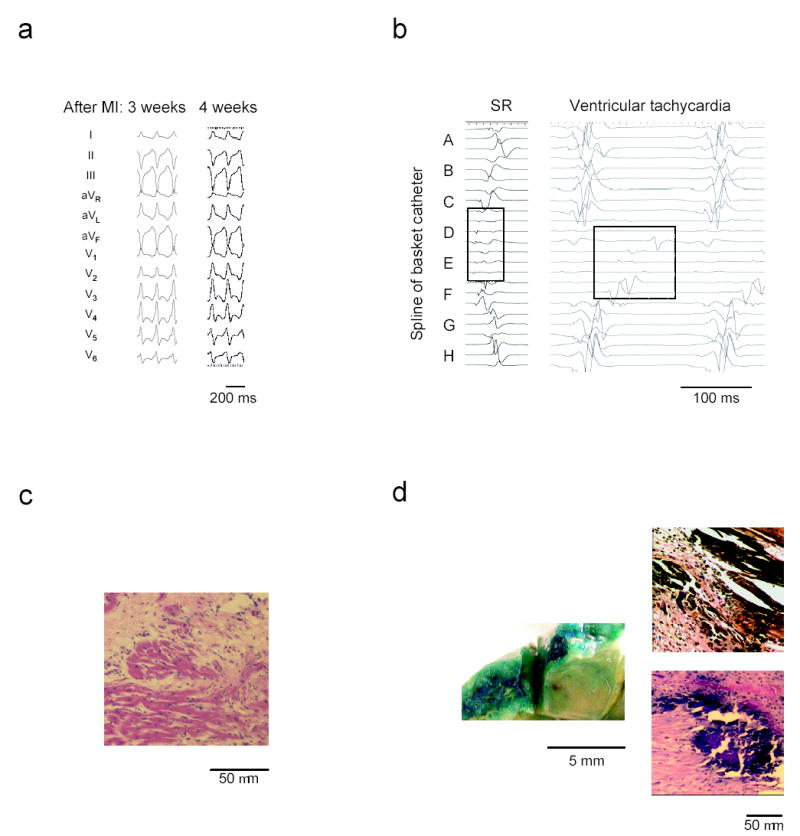
Ventricular tachycardia after myocardial infarction in a porcine model.
(a) ECG example showing identical VT morphology and cycle
length from one week to the next in a representative animal.
(b) Intracardiac electrograms during sinus rhythm (SR) show low
amplitude, fractionated signals in the anterior septal region (splines D and
E). The ventricular tachycardia recording demonstrates slow, progressive
activation of this region through diastole. (c)
Hematoxylin-Eosin stained microsection of the infarct border zone reveals
surviving strands of myocardium surrounded by fibrotic scar in the anterior
septum. (d) X-gal staining to identify lacZ
gene transfer. (left) Gross tissue shows intense blue staining indicative of
lacZ expression in the target area at the anterior
septal border zone. (right) Microscopic sections taken from the target
region exhibit blue, lacZ positive myocytes.
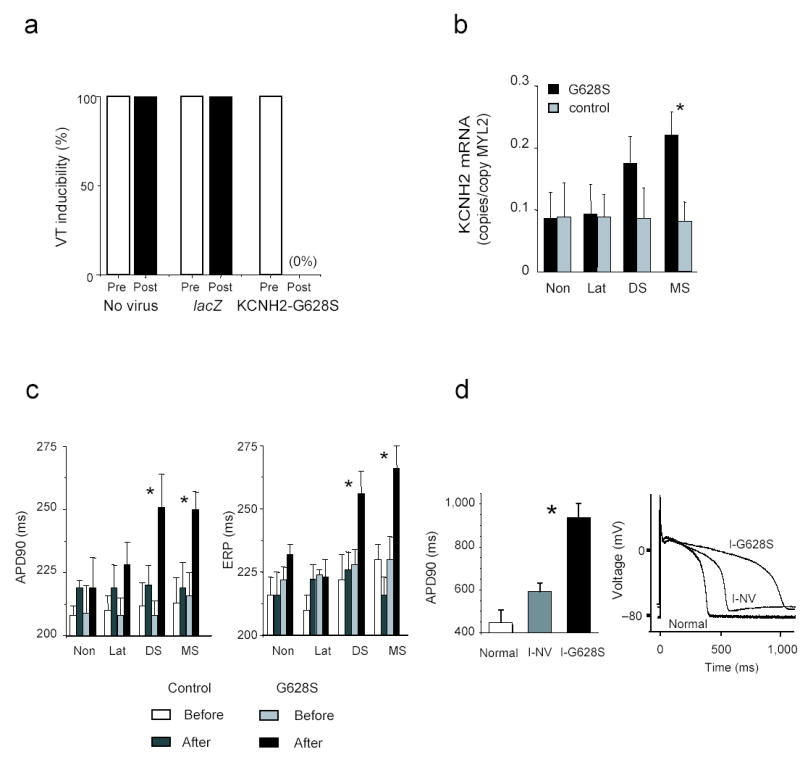
KCNH2-G628S gene transfer in the MI-VT model.
(a) Prior to gene transfer, VT was repeatedly inducible in all
animals. After gene transfer, no arrhythmias could be induced in the
KCNH2-G628S infected animals. All control animals
remained inducible. (b) RT-PCR results show increased ERG mRNA
in mid and distal septum of G628S animals. Data is normalized to MCL-2v
expression (copies of ERG per copy of MLC2v). Non—non-infarcted
basal lateral wall. MS—mid-anterior septal infarct border.
DS—distal anterior septal infarct border.
Lat—anterior lateral infarct border. (c)
Measurement of action potential duration to 90% repolarization
(APD90) and effective refractory period (ERP) from the indicated regions.
Prolongation of APD90 and ERP is isolated to the septal border of the
infarct. (d) Patch clamp data of action potentials from
uninfarcted anterior septal cells (Normal) compared to infarct-no virus
(I-NV) or infarct-KCNH2-G628S (I-G628S) infected cells from
the anterior septal infarct border zone. The left panel shows summary data
from the indicated groups. The right panel shows representative tracings.
* p < 0.05.
Similar articles
-
Gene therapy approaches to ventricular tachyarrhythmias.J Electrocardiol. 2007 Nov-Dec;40(6 Suppl):S187-91. doi: 10.1016/j.jelectrocard.2007.05.036. J Electrocardiol. 2007. PMID: 17993320 Free PMC article.
-
Ventricular tachycardia from the healed myocardial infarction scar: validation of an animal model and utility of gene therapy.Heart Rhythm. 2009 Aug;6(8 Suppl):S91-7. doi: 10.1016/j.hrthm.2009.03.048. Epub 2009 Apr 1. Heart Rhythm. 2009. PMID: 19631912 Free PMC article.
-
Short QT syndrome. What is it? Where is it?Indian Heart J. 2005 Nov-Dec;57(6):769-71. Indian Heart J. 2005. PMID: 16521657 Review. No abstract available.
-
Selective molecular potassium channel blockade prevents atrial fibrillation.Circulation. 2010 Jun 1;121(21):2263-70. doi: 10.1161/CIRCULATIONAHA.109.911156. Epub 2010 May 17. Circulation. 2010. PMID: 20479154 Free PMC article.
-
Molecular genetic basis of sudden cardiac death.Pediatr Clin North Am. 2004 Oct;51(5):1229-55. doi: 10.1016/j.pcl.2004.04.012. Pediatr Clin North Am. 2004. PMID: 15331282 Review.
Cited by
-
Heterogeneous repolarization creates ventricular tachycardia circuits in healed myocardial infarction scar.Nat Commun. 2022 Feb 11;13(1):830. doi: 10.1038/s41467-022-28418-1. Nat Commun. 2022. PMID: 35149693 Free PMC article.
-
Allogeneic cardiospheres delivered via percutaneous transendocardial injection increase viable myocardium, decrease scar size, and attenuate cardiac dilatation in porcine ischemic cardiomyopathy.PLoS One. 2014 Dec 2;9(12):e113805. doi: 10.1371/journal.pone.0113805. eCollection 2014. PLoS One. 2014. PMID: 25460005 Free PMC article.
-
Tachycardia in post-infarction hearts: insights from 3D image-based ventricular models.PLoS One. 2013 Jul 2;8(7):e68872. doi: 10.1371/journal.pone.0068872. Print 2013. PLoS One. 2013. PMID: 23844245 Free PMC article.
-
Gene Therapy for Cardiac Arrhythmias.Acta Cardiol Sin. 2013 May;29(3):226-34. Acta Cardiol Sin. 2013. PMID: 27122711 Free PMC article. Review.
-
Targeted high-efficiency, homogeneous myocardial gene transfer.J Mol Cell Cardiol. 2007 May;42(5):954-61. doi: 10.1016/j.yjmcc.2007.02.004. Epub 2007 Feb 14. J Mol Cell Cardiol. 2007. PMID: 17484913 Free PMC article.
References
-
- American Heart Association, 2001. Heart and Stroke Statistical Update. American Heart Association; Dallas, TX: 2002.
-
- Rothman S, et al. Circulation. 1997;96:3499–3508. - PubMed
-
- Gould P, Krahn A Canadian Heart Rhythm Society Working Group on Device Advisories. JAMA. 2006;295:1907–1911. - PubMed
-
- Maisel W, et al. JAMA. 2006;295:1901–1906. - PubMed
Publication types
MeSH terms
Substances
Grants and funding
LinkOut - more resources
Full Text Sources
Other Literature Sources
Medical
