. 2006 Sep;12(9):1353-60.
doi: 10.3201/eid1209.060276.

Genomic signatures of human versus avian influenza A viruses

Affiliations

Genomic signatures of human versus avian influenza A viruses

Guang-Wu Chen et al. Emerg Infect Dis. 2006 Sep.

Abstract

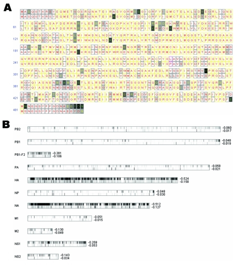
Position-specific entropy profiles created from scanning 306 human and 95 avian influenza A viral genomes showed that 228 of 4591 amino acid residues yielded significant differences between these 2 viruses. We subsequently used 15,785 protein sequences from the National Center for Biotechnology Information (NCBI) to assess the robustness of these signatures and obtained 52 "species-associated" positions. Specific mutations on those points may enable an avian influenza virus to become a human virus. Many of these signatures are found in NP, PA, and PB2 genes (viral ribonucleoproteins [RNPs]) and are mostly located in the functional domains related to RNP-RNP interactions that are important for viral replication. Upon inspecting 21 human-isolated avian influenza viral genomes from NCBI, we found 19 that exhibited > or =1 species-associated residue changes; 7 of them contained > or =2 substitutions. Histograms based on pairwise sequence comparison showed that NP disjointed most between human and avian influenza viruses, followed by PA and PB2.


Figures

Figure
Figure
A) Entropy plot for avian versus human influenza viruses for NP amino acid residues. In each aligned position, we have a consensus residue for 95 avian strains displayed on top and a consensus residue for 306 human strains at the bottom. Completely conserved amino acid positions are filled with white; less conserved amino acids are filled in various gray shadings. Positions in which 1 single residue dominates >90%, <90% but >75%, and <75% are labeled with red, yellow, and green letters, respectively. Yellow rectangles indicate that both human and avian viruses are completely conserved to the same residue; magenta rectangles indicate that avian and human viruses are each completely conserved to a different residue. B) Entropy plots for the entire influenza A viral genome. Each lane displays entropy value distributions of aligned protein sequences for 1 of the 11 viral proteins; the upper half represents 95 avian strains, and the bottom half represents 306 human strains. (PB1-F2 contains fewer strains, as described in Discussion.) Positions completely conserved to a single residue are shown in a white band, while less conserved ones are shown in various gray shadings. The average entropy for the entire segment is shown to the right of these lanes. Entropy values are zero when residues are completely conserved; more negative values indicate more diversity. Alignment size for each protein from top to bottom is 759, 757, 90, 716, 591, 498, 480, 252, 97, 230, and 121.
Figure A1
Figure A1
Entropy plot for all 11 influenza proteins for human (top) versus avian (bottom). In each aligned position, we have a consensus residue for 95 avian strains displayed on top, and a consensus residue for 306 human strains at the bottom. Completely conserved amino acid positions are filled with white, while less conserved amino acids are filled in various gray shadings. Positions where one single residue dominates over 90%, less than 90% but greater than 75%, and less than 75% are labeled with red, yellow, and green letters, respectively. Yellow rectangles indicate that both human and avian flu are completely conserved to the same residue, while rectangles in magenta indicate that avian and human flu each completely conserves to a different residue Additional plots for HA, NA, NS1 and NS2, for using different counts of human or avian strains are detailed as individual captions to these plots. Adobe Acrobat PDF available at http://wwwnc.cdc.gov/eid/pdfs/06-0276-FA1.pdf (21 pages).
Figure A2
Figure A2
Histograms on comparing 306 human versus 95 avian influenza A viruses, based on nucleotide pairwise sequence identities. Vertical axis shows the count for pairs of sequences with specific percent identity (rounded to integer). Red bars represent frequencies for 'homo' pairs – sequences of the same host species (human to human, or avian to avian); blue bars represent frequencies for 'hetero' pairs – pairs that cross host species (human to avian, or avian to human). Adobe Acrobat PDF available at http://wwwnc.cdc.gov/eid/pdfs/06-0276-FA2.pdf (6 pages).
Figure A3
Figure A3
Histograms compare 43 avian allele A viruses and 306 human viruses (panels A and C), and 52 avian allele B viruses and 306 human viruses (panels B and D), based on their NS1 and NS2 genomic segments. Vertical axis shows the count for pairs of sequences with specific percent identity (rounded to integer). Red bars represent frequencies for 'homo' pairs – sequences of the same host species (human to human, or avian to avian); blue bars represent frequencies for 'hetero' pairs – pairs that cross host species (human to avian, or avian to human). Adobe Acrobat PDF available at http://wwwnc.cdc.gov/eid/pdfs/06-0276-FA3.pdf (5 pages).

References

    1. Scholtissek C, Rohde W, von Hoyningen V, Rott R. On the origin of the human influenza virus subtypes H2N2 and H3N2. Virology. 1978;87:13–20. 10.1016/0042-6822(78)90153-8 - DOI - PubMed
    1. Reid AH, Taubenberger JK, Fanning TG. Evidence of an absence: the genetic origins of the 1918 pandemic influenza virus. Nat Rev Microbiol. 2004;2:909–14. 10.1038/nrmicro1027 - DOI - PMC - PubMed
    1. Taubenberger JK, Reid AH, Lourens RM, Wang R, Jin G, Fanning TG. Characterization of the 1918 influenza virus polymerase genes. Nature. 2005;437:889–93. 10.1038/nature04230 - DOI - PubMed
    1. Chang SC, Cheng YY, Shih SR. Avian influenza virus: the threat of a pandemic. Chang Gung Med J. 2006;29:130–4. - PubMed
    1. Rice P, Longden I, Bleasby A. EMBOSS: the European Molecular Biology Open Software Suite. Trends Genet. 2000;16:276–7. 10.1016/S0168-9525(00)02024-2 - DOI - PubMed

Publication types

MeSH terms

Associated data