. 2007 Jan;27(2):411-25.
doi: 10.1128/MCB.01391-06. Epub 2006 Oct 30.

A cooperative action of the ATP-dependent import motor complex and the inner membrane potential drives mitochondrial preprotein import

Affiliations

A cooperative action of the ATP-dependent import motor complex and the inner membrane potential drives mitochondrial preprotein import

Martin Krayl et al. Mol Cell Biol. 2007 Jan.

Abstract

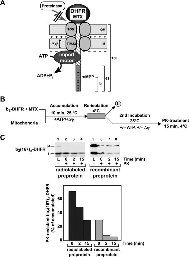
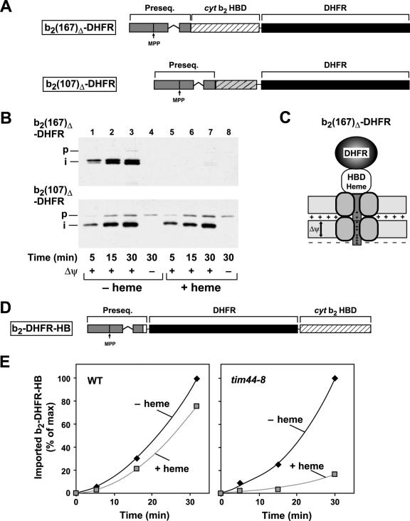
The import of mitochondrial preproteins requires an electric potential across the inner membrane and the hydrolysis of ATP in the matrix. We assessed the contributions of the two energy sources to the translocation driving force responsible for movement of the polypeptide chain through the translocation channel and the unfolding of preprotein domains. The import-driving activity was directly analyzed by the determination of the protease resistances of saturating amounts of membrane-spanning translocation intermediates. The ability to generate a strong translocation-driving force was solely dependent on the activity of the ATP-dependent import motor complex in the matrix. For a sustained import-driving activity on the preprotein in transit, an unstructured N-terminal segment of more than 70 to 80 amino acid residues was required. The electric potential of the inner membrane was required to maintain the import-driving activity at a high level. The electrophoretic force of the potential exhibited only a limited capacity to unfold preprotein domains. We conclude that the membrane potential increases the probability of a dynamic interaction of the preprotein with the import motor. Polypeptide translocation and unfolding are mainly driven by the inward-directed translocation activity based on the functional cooperation of the import motor components.


Figures

FIG. 1.
FIG. 1.
Assay used to determine the inward-directed translocation force in mitochondria. (A) Schematic drawing of the translocation-driving activity assay. The MTX-stabilized DHFR domain blocks the import of the preprotein b2(167)Δ-DHFR. The unstructured N-terminal segment of the preprotein is inserted into the import channel and can interact with the import motor if it crosses both mitochondrial membranes. The generation of an inward-directed translocation force prevents the degradation of the preprotein in transit by external proteases. The MPP cleaves off the first 31 amino acids in the presequence of cytochrome b2 as indicated. The locations of the positively charged amino acids in the presequence are indicated (+). (B) Diagram of the experimental procedure. The cytochrome b2-DHFR fusion proteins are preincubated with the specific ligand MTX. In the presence of ATP and the membrane potential, the preprotein in its MTX-stabilized state is bound to the mitochondria. Preproteins that are not inserted in the import channel are removed by a reisolation step. Subsequently, the load sample (L) is taken and the matrix ATP or inner Δψ is depleted or sustained. Samples are taken over time and subsequently treated with proteinase K. (C) Protease resistance of b2(167)Δ-DHFR translocation intermediates. Radiolabeled preproteins generated by in vitro translation and purified recombinant preproteins were stabilized by MTX and incubated with wild-type mitochondria (p, precursor; i, processing intermediate). PK was added at the indicated time points in the absence of a Δψ. The amount of the remaining fusion proteins bound to mitochondria was determined by autoradiography or by Western blotting, using antibodies against DHFR. The value obtained for the accumulated i-b2(167)Δ-DHFR (L) in the absence of PK was set to 100%.
FIG. 2.
FIG. 2.
Matrix ATP levels and conditional mutants of the import motor complex determine the protease resistance of translocation intermediates. Experiments and quantifications were performed as described in the legend to Fig. 1. The reactions were performed with conditional-mutant mitochondria from the ssc1-2 (A), tim44-8 (B), and pam16-1 (C) strains. The temperature-sensitive phenotype was induced by incubation of the isolated mitochondria for 15 min at 37°C prior to the import reaction. The protease-resistant processed form of b2(167)Δ-DHFR was quantified, and the amount obtained in the absence of protease was set to 100%.
FIG. 3.
FIG. 3.
The length of the unstructured segment traversing both mitochondrial membranes is critical for the generation of an inward-directed translocation force. (A) Schematic graph of MTX-bound translocation intermediates of the preproteins b2(107)Δ-DHFR, b2(98)Δ-DHFR, and b2(84)Δ-DHFR traversing the mitochondrial membranes. Approximately 50 amino acids in an unstructured state are required to span both membranes. (B) The assay for an import-driving activity was performed as described in the legend to Fig. 1 with the cytochrome b2-DHFR fusion proteins shown in panel A. (C) Mitochondria from the wild-type (WT) and the temperature-sensitive mutant ssc1-2 and ssc1-3 strains were incubated with recombinant b2(167)Δ-DHFR in the presence or absence of MTX as described in Materials and Methods. Mitochondria were reisolated, lysed under native conditions, and analyzed by BN-PAGE. The translocase complexes were visualized by Western blotting and immunodecoration with antibodies directed against Tim23. Indicated are the molecular masses and the localizations of the protein complexes. (D) The formation of TOM-TIM supercomplexes was analyzed as described above using DHFR fusion proteins with the indicated length of the presequence and mature part derived from cytochrome b2.
FIG. 4.
FIG. 4.
The Δψ assists the generation of an inward-directed translocation force. (A) The assay for an import-driving activity and its quantification were performed as described in the legend to Fig. 1 using the preprotein b2(167)Δ-DHFR dependent on the Δψ. The matrix ATP levels were reduced by treatment with apyrase (−ATP) as indicated. The membrane potential was depleted by the addition of valinomycin, antimycin, and oligomycin as indicated. (B) Time course of protease resistance after depletion of the membrane potential. The experiment was performed as described in the legend to Fig. 1. The Δψ was depleted by the addition of valinomycin at the indicated time points. (C) Backward release of preproteins from the translocation channel is not affected by import motor mutants. The radiolabeled preprotein Su9(86)-DHFR was accumulated as an MTX translocation intermediate in wild-type and ssc1-3 mutant mitochondria under nonpermissive conditions. Mitochondria were reisolated, and the membrane potential was depleted as indicated. The amount of precursor associated with the mitochondria was monitored over the indicated incubation times by SDS-PAGE of the mitochondrial pellet and detection by autoradiography.
FIG. 5.
FIG. 5.
The rate and efficiency of mitochondrial import are dependent on the lengths of the membrane-spanning segments in front of folded domains. (A) Import of recombinant purified cytochrome b2-DHFR fusion proteins b2(167)Δ-DHFR, b2(84)Δ-DHFR, and b2(47)-DHFR in native and urea-denatured states. The import reaction was performed as described in Materials and Methods (p, precursor; i, processing intermediate). Nonimported preprotein was removed by treatment with proteinase K. The proteins were separated by SDS-PAGE and detected by Western blotting and immunodecoration with antibodies against DHFR. Import of the recombinant purified fusion proteins b2(167)Δ-DHFR (B) and b2(47)-DHFR (C) was performed dependent on the membrane potential. The proteins were imported in their native or urea-denatured state in the presence of increasing amounts of the ionophore CCCP. The imported and processed preproteins were quantified, and the value obtained for the control (0 μM CCCP) was set to 100%. (D) The assay for an import-driving activity for the fusion proteins b2(167)Δ-DHFR and b2(47)-DHFR was performed as described in the legend to Fig. 1. (E) Schematic graph of an MTX-bound translocation intermediate of b2(47)-DHFR inserted into the mitochondrial membranes.
FIG. 6.
FIG. 6.
The unfolding of the stable folded HBDs cannot be initiated by the membrane potential alone. (A) Schematic representation of the preproteins b2(167)Δ-DHFR and b2(107)Δ-DHFR. The presequence, the HBD of cytochrome b2, and the DHFR domain are labeled. (B) The import of the recombinant purified preproteins b2(167)Δ-DHFR and b2(107)Δ-DHFR was performed as described in Material and Methods in the absence or presence of heme. Nonimported proteins were removed by proteinase K. Proteins were separated by SDS-PAGE and detected by Western blotting and immunodecoration with antibodies against DHFR. (C) Schematic representation of the preprotein b2(167)Δ-DHFR with a stably folded HBD traversing both mitochondrial membranes. (D) Schematic representation of the preprotein b2-DHFR-HB. The fusion protein consists of the presequence of cytochrome (cyt) b2, the DHFR domain, and the HBD of cytochrome b2 as indicated. (E) The recombinant purified preprotein b2-DHFR-HB was imported into wild-type and tim44-8 mitochondria under nonpermissive conditions in the absence and presence of heme. The imported and processed protein was quantified. The maximal amount of protein imported was set to 100%.

Similar articles

Cited by

References

    1. Bauer, M. F., S. Hofmann, W. Neupert, and M. Brunner. 2000. Protein translocation into mitochondria: the role of TIM complexes. Trends Cell Biol. 10:25-31. - PubMed
    1. Bauer, M. F., C. Sirrenberg, W. Neupert, and M. Brunner. 1996. Role of Tim23 as voltage sensor and presequence receptor in protein import into mitochondria. Cell 87:33-41. - PubMed
    1. Bömer, U., A. C. Maarse, F. Martin, A. Geissler, A. Merlin, B. Schönfisch, M. Meijer, N. Pfanner, and J. Rassow. 1998. Separation of structural and dynamic functions of the mitochondrial translocase: Tim44 is crucial for the inner membrane import sites in translocation of tightly folded domains, but not of loosely folded preproteins. EMBO J. 17:4226-4237. - PMC - PubMed
    1. Chacinska, A., P. Rehling, B. Guiard, A. E. Frazier, A. Schulze-Specking, N. Pfanner, W. Voos, and C. Meisinger. 2003. Mitochondrial translocation contact sites: separation of dynamic and stabilizing elements in formation of a TOM-TIM-preprotein supercomplex. EMBO J. 22:5370-5381. - PMC - PubMed
    1. Craig, E. A., J. Kramer, J. Shilling, M. Werner-Washburne, S. Holmes, J. Kosic-Smithers, and C. M. Nicolet. 1989. SSC1, an essential member of the yeast HSP70 multigene family, encodes a mitochondrial protein. Mol. Cell. Biol. 9:3000-3008. - PMC - PubMed

Publication types

MeSH terms

Substances

LinkOut - more resources